(How to Program) Paul Deitel, Harvey Deitel-Java How to Program, Early Object...
Long Mac Kinnon05.Dl.pdf
1. RESEARCH ARTICLES
laboratory for helpful discussions; R. Jain for initial
49. K. McCormack, T. McCormack, M. Tanouye, B. Rudy, coordinates and structure factors have been deposited
experiments with lipids; Q. Wang and B. T. Chait for
W. Stuhmer, FEBS Lett. 370, 32 (1995). with the Protein Data Bank with accession ID 2A79.
mass spectrometry; R. Dutzler for assistance with data
50. G. Shi et al., Neuron 16, 843 (1996).
collection; O. Pongs for Kv1.2 DNA; J. Trimmer for b2
51. N. Nagaya, D. M. Papazian, J. Biol. Chem. 272, 3022 Supporting Online Material
(1997). www.sciencemag.org/cgi/content/full/1116269/DC1
subunit DNA; Brookhaven National Laboratory (Na-
52. T. A. Jones, J. Y. Zou, S. W. Cowan, M. Kjeldgaard, Materials and Methods
tional Synchrotron Light Source beamlines X25 and
Acta Crystallogr. A47, 110 (1991). References
X29) and the Swiss Light Source (beamline PX1) staff
53. M. Carson, Methods Enzymol. 277, 493 (1997). for assistance in data collection; and W. Chin for help
54. P. Kraulis, J. Appl. Crystallogr. 24, 946 (1991). 17 June 2005; accepted 5 July 2005
with manuscript preparation. This work was supported
55. A. Nicholls, K. A. Sharp, B. Honig, Proteins 11, 281 Published online 7 July 2005;
in part by NIH grant no. GM43949 to R.M. and NIH
(1991). 10.1126/science.1116269
grant no. RR00862 to B. T. Chait. R.M. is an Investigator
56. We thank A. Lee, V. Ruta, and members of the MacKinnon Include this information when citing this paper.
in the Howard Hughes Medical Institute. Atomic
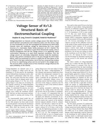
Voltage Sensor of Kv1.2: How and to what extent S4 moves has been
the subject of much debate. Gating-dependent
reactivity of sulfhydryl reagents with cysteine
Structural Basis of residues led to an initial hypothesis of large
(È15 )) translations of S4 in some models
Electromechanical Coupling (13–17). But very small distance changes
measured in fluorescence resonance energy
transfer (FRET) experiments suggested much
Stephen B. Long, Ernest B. Campbell, Roderick MacKinnon*
Downloaded from www.sciencemag.org on October 10, 2007
smaller movements of S4 across the membrane
(18, 19). Crevices surrounding S4 were in-
Voltage-dependent ion channels contain voltage sensors that allow them to
voked to account for the sulfhydryl reactivity
switch between nonconductive and conductive states over the narrow range
in the setting of these smaller movements, with
of a few hundredths of a volt. We investigated the mechanism by which these
translations and/or rotations of S4 occurring
channels sense cell membrane voltage by determining the x-ray crystal
across a narrow neck inside an hourglass-
structure of a mammalian Shaker family potassium ion (Kþ) channel. The
shaped canaliculus. In the transporter model
voltage-dependent Kþ channel Kv1.2 grew three-dimensional crystals, with an
hypothesis, the S4 does not change its depth
internal arrangement that left the voltage sensors in an apparently native
in the membrane at all (less than 3 ) move-
conformation, allowing us to reach three important conclusions. First, the
ment) (18, 20). Instead, the field is moved
voltage sensors are essentially independent domains inside the membrane.
over the S4 charges by alternately opening and
Second, they perform mechanical work on the pore through the S4-S5 linker
closing crevices to the internal and external
helices, which are positioned to constrict or dilate the S6 inner helices of the
solutions.
pore. Third, in the open conformation, two of the four conserved Arg residues
The above models of voltage-dependent
on S4 are on a lipid-facing surface and two are buried in the voltage sensor.
gating vary in detail, but they have had two
The structure offers a simple picture of how membrane voltage influences the
essential features in common. First, the S4
open probability of the channel.
helix is sequestered from the lipid membrane
Ealthough Larsson and colleagues proposed that
gating charges move; the main subject of study
Voltage-dependent ion channels open in re-
has been the Shaker voltage-dependent Kþ
sponse to changes in voltage across the cell a surface of S4 could be exposed to lipid (11)^.
membrane (1). In this process, the membrane Second, the voltage-sensor helices S1 to S4 are
(Kv) channel, and numerous models have been
electric field performs mechanical work to alter packed tightly against the a helices of the pore.
put forth. One fundamental constraint for any
model is that when a Shaker Kþ channel
the channel_s conformation within the mem- In other words, it was reasonably assumed that
brane. The work arises from the force exerted voltage-dependent ion channels are like con-
opens, it transfers the net equivalent of 12 to
by the electric field on charged amino acids, ventional a-helical membrane proteins that
14 positive elementary charges across the
termed gating charges (1–3). The size of the membrane electric field from inside to outside, form a fairly rigid disk of helices in the mem-
gating charge is very large (4), accounting for brane. A first hint that a rigid disk of helices
and most of this charge is carried by four S4
the exquisite sensitivity of voltage-dependent Arg residues on each of four identical channel might not pertain to voltage-dependent ion
ion channels to small changes in membrane channels came from the demonstration that the
subunits (4–6).
voltage. To understand this process, one must A guiding assumption underlying most voltage sensor (S1 to S4) from the Shaker Kv
first answer two questions: How do gating channel could be spliced onto the pore of
models for the voltage sensor has been that
KcsA (a non–voltage-dependent Kþ channel)
charges move within the membrane electric the S4 helix with its Arg residues is completely
field? And how are these movements mechan- (7–10), or mostly (11), sequestered from the to confer voltage-dependent gating (21). This
ically coupled to opening and closing of the membrane, in order to protect the charges from finding implied that the voltage sensor might
pore? the lipid_s low dielectric environment. To ac- be an almost-independent domain, because if it
No experimentally based model has yet complish this, most models postulate that the had to form a large interface through helix
provided answers to both of these questions. S4 helix inserts into a groove at the interface packing with the pore, the chimera would
between adjacent subunits of the Kþ channel
So far, little progress has been made toward likely not function.
the second question concerning the mechanical The first atomic structures of a prokaryotic
tetramer, such that pore a helices S5 and S6
coupling of voltage-sensor movements to the form a wall on one side of S4 and voltage- Kv channel (KvAP) also implied that the
pore. Most effort has focused on how the voltage sensors are loosely attached to the pore
sensor a helices S1 to S3 form a wall on the
(22). One of these EProtein Data Bank (PDB)
other side, the lipid-facing perimeter, to create
Howard Hughes Medical Institute, Laboratory of
ID 1ORS^ was of an isolated voltage-sensor
a gating channel or protein-lined canaliculus
Molecular Neurobiology and Biophysics, Rockefeller
for S4 (7–10, 12). This arrangement would domain, which surprisingly could be expressed
University, 1230 York Avenue, New York, NY 10021,
in the membrane by itself (without the pore).
allow the S4 helix to move its charged amino
USA.
acids across the membrane without exposing Another, which is a full-length channel struc-
*To whom correspondence should be addressed.
ture (PDB ID 1ORQ), showed the voltage-
them to the lipid environment.
E-mail: mackinn@rockefeller.edu
903
www.sciencemag.org SCIENCE VOL 309 5 AUGUST 2005
2. RESEARCH ARTICLES
sensor domains in a non-native conformation, that some of the Arg residues move more than itive information on the mechanism of voltage-
15 ) through the thickness of the membrane dependent gating.
pulled toward the cytoplasmic side of the pore.
Voltage-sensor structure: Relationship
Not only had the voltage sensors undergone (23, 24).
between Kv1.2 and KvAP. Electron density
domain-like movements with respect to the The KvAP studies implied that the voltage
pore, but the sensors seemed to have a great sensors are highly mobile; that S4 is not in a maps are weak in the region of the voltage
deal of internal flexibility. This was unusual canaliculus; and possibly that some S4 Arg sensor, relative to the remainder of the chan-
nel. However, the four transmembrane helices
behavior for a membrane protein. The Arg- residues could be exposed to the lipid mem-
(S1 to S4) were easily recognizable, with side-
containing S4 helix formed part of a helix-turn- brane, which would allow the voltage-sensing
chain density for some, but not all, amino acids
helix structure (termed a voltage-sensor paddle) apparatus to exploit opposing electrostatic and
(26). Electron density was present for the first
through its antiparallel relationship to S3b (the hydrophobic forces to gate the channel (23, 25).
(Arg294), second (Arg297), and third (Arg300)
C-terminal half of S3). The paddle was pro- However, a major weakness of the KvAP studies
posed to move at the protein-lipid interface was directly related to the voltage sensors_ Arg residues on S4, and for two Phe residues
with S3b Babove[ and S4 Bbelow.[ Experi- (Phe302 and Phe305), establishing the correct
mobility: Distortions associated with extraction
ments with avidin capture of biotin suggested from the membrane left many aspects of the register of this helix (Fig. 1). The partial model
structure uncertain. Most notably, the connec- of the voltage sensor contains helices for S1
tions between the voltage sensors and pore were (19 amino acids), S2 (residues 219 to 243), S3
disrupted. The crystals of Kv1.2 have maintained (21 amino acids), S4 (residues 288 to 311), and
these connections and thus convey more defin- the S4-S5 linker (residues 312 to 325). Com-
Downloaded from www.sciencemag.org on October 10, 2007
Fig. 1. Density for S4 and the S4-S5 linker
calculated without a voltage-sensor model. The
electron density map for S4 and the S4-S5 linker
(blue mesh) is shown with the final model drawn
as a Ca trace (gray) with side-chain residues
(yellow, blue, and red sticks). Arg (R) residues 1
to 3 on S4 have density for their side chains.
Density for these side chains and for two Phe
residues (F302 and F305) helped establish the
correct register of the S4 helix. The side chain of
Arg 4 is truncated after the Cb atom (modeled as
alanine) because density past this point was not
present in the maps. A few other residues in S4
have also been modeled as alanine. Phases for Fig. 2. Stereoviews comparing the Kv1.2 structure with two structures of the prokaryotic Kv channel
the map were calculated by removing the entire KvAP. (A) A single subunit of the integral membrane pore and partial model of the voltage sensor of
voltage sensor (S1 to S4 helices through residue Kv1.2 viewed from the side as a gray Ca trace. Arg residues 1 to 4 on the S4 helix (blue labels) are
313 of the S4-S5 linker) from the model and depicted as yellow and blue sticks. The side chain for Arg 4, although not included in the final
refining the remaining partial structure of the coordinates, is modeled in a chemically reasonable conformation for the purpose of illustration. (B) The
pore and T1/b complex using a simulated Kv1.2 structure (gray) viewed as in (A) with a full-length crystal structure of KvAP (red Ca trace, PDB ID
annealing protocol in the CNS software (41). 2A0L) superimposed by alignment of main-chain atoms of the pore helices and selectivity filter, and
This procedure, which is used to generate a with an isolated voltage-sensor structure of KvAP (Aeropyrum Pernix Voltage Sensor, or APVS) (blue
‘‘simulated annealing omit map,’’ essentially Ca trace, PDB ID 1ORS) (22) superimposed by alignment of main-chain atoms of a helices S1 and S2.
eliminates bias in the map. The map is a 2Fo-Fc (C) A hypothetical model of a single KvAP subunit is shown as a red Ca trace with yellow and blue side
map (where Fo is the observed structure factor chains for Arg residues 1 to 4 on the S4 helix. This was constructed by combining the isolated voltage
and Fc is the calculated structure factor) that was sensor and pore of KvAP according to their positions relative to Kv1.2 as displayed in (B). The S4-S5
˚
calculated from 30 to 2.9 A, contoured at 0.5s, linker residues of KvAP (residues 136 to 146) are positioned relative to the pore and voltage sensor
based on the Kv1.2 S4-S5 helix. A queue of Kþ ions (green spheres) from the pore are shown as a
and drawn around the portion of the molecule
shown. M, Met; G, Gly. reference in (A) to (C). The figure was generated with Molscript software (42).
904 5 AUGUST 2005 VOL 309 SCIENCE www.sciencemag.org
3. RESEARCH ARTICLES
Kv1.2, the presence of a T1 domain and its con-
parisons with KvAP crystal structures assisted 2A0L, red) is aligned with the pore of Kv1.2,
nection to the S1 helix undoubtedly help to main-
in the identification of these helices. Most side and an isolated voltage-sensor structure of
tain a native conformation of the voltage sensor.
chains were included on S4, the S4-S5 linker, KvAP [PDB ID 1ORS, blue (22)] is aligned
We conclude that the basic architecture of
and S2. S1 and S3 were built with alanine with the voltage sensor of Kv1.2. A model of
the voltage sensor in a membrane is similar in
residues. Loops connecting helices S1 to S2 KvAP can be made to look like Kv1.2 by
Kv1.2 and KvAP. They both have an antipar-
and S3 to S4 were omitted because electron simply repositioning its voltage sensor to more
allel arrangement of S3 and S4. This arrange-
density was weak or absent. The turn connect- closely resemble the isolated voltage-sensor
ment was called a voltage-sensor paddle in
ing S2 to S3, which varies in its conformation structure (Fig. 2C). The linker connecting the
KvAP (22). In KvAP, there are two distinct
in different crystal structures of KvAP (22) voltage sensor to the pore in KvAP is the
segments of S3, termed S3a and S3b. In
(and see PDB ID 2A0L), was also omitted. appropriate length and has the correct amphi-
Kv1.2, the electron density for S3 appears to
Thus, this partial model of the voltage sensor is pathicity (see below) to match the linker in
be a single helix with a bend, presumably near
missing several elements, but it still addresses Kv1.2. Apparently, upon extraction of KvAP
the connection between S3a and S3b. This
many important questions. from the lipid membrane, the voltage sensor is
finding probably represents a different posi-
A model of a single subunit of Kv1.2 is dislodged from its proper position. This kind
tioning of the paddle from that which we
shown in Fig. 2A. Two different crystal struc- of distortion in multiple crystal structures of
observed in KvAP. Here we do not distinguish
tures of KvAP superimposed on Kv1.2 show KvAP (22) (and see PDB ID 2A0L) probably
between S3a and S3b in Kv1.2, but simply
the relationship between these channels (Fig. reflects the importance of a cell membrane to
refer to the antiparallel unit formed by the S3
2B). A full-length KvAP structure (PDB ID hold the voltage sensor in its proper position. In
and S4 helices as the voltage-sensor paddle.
Downloaded from www.sciencemag.org on October 10, 2007
The comparison of Kv1.2 and KvAP serves
many useful purposes. First, their fundamental
similarity reinforces our confidence in the
accuracy of the Kv1.2 voltage-sensor model
and shows that S4 and S3 form a voltage-
sensor paddle as in KvAP. Second, because
Kv1.2 and KvAP are similar, we can consider
KvAP functional data in constraining possible
motions of the Kv1.2 voltage sensor. Third,
certain differences between their structures
may provide useful information about move-
ments of the voltage sensor. For example, the
voltage-sensor paddles in Kv1.2 are in a
slightly different position with respect to the
S1 and S2 helices (Fig. 2B). This is under-
standable if the paddles are mobile, allowing
them to move in the gating process. Fourth, S4
(and S3) is nearly two helical turns longer at its
extracellular end in Kv1.2 than in KvAP (the
Kv1.2 S4 contains two extra helical turns pre-
ceding the Arg residues) (compare Fig. 2, A
and C). This means the paddle in Kv1.2 will
project further into the extracellular solution
and therefore may exhibit differences (relative
to KvAP) in accessibility to spider toxins and
small molecules that interact with the voltage
sensor from outside the cell.
Voltage-sensor coupling to the pore:
The S4-S5 linker helix. The S4-S5 linker is
an amphipathic a helix that runs parallel to the
membrane plane inside the cell, with its hy-
Fig. 3. The connection between the voltage sensor and the pore in the Kv1.2 channel. (A) The S6 inner
drophobic surface facing the membrane and its
helix (residues 388 to 421) is shown as a gray and green ribbon with yellow side chains for Pro-Val-Pro
polar surface facing the cytoplasm (Figs. 2A
(residues 405 to 407), and the S4-S5 linker and S5 from the same subunit (residues 311 to 342) are
and 3A). The most important aspect of this
shown as a gray and green ribbon. Side chains on the S4-S5 linker are yellow (carbon), red (oxygen),
and blue (nitrogen). The perspective is from the side of the channel near the intracellular water helix is its position against the pore; it crosses
(below)/membrane (above) interface. Regions colored green were necessary to transfer the Shaker over the top of the S6 inner helix from the
voltage sensor to KcsA (32). (B) Residues on the S4-S5 linker in direct contact with residues on S6 are
same subunit and makes many amino acid
shown as red and blue spheres, respectively. The helices are drawn as ribbons and colored in the
contacts with it (Fig. 3, A and B). The S6 inner
following manner: S4-S5, red; S5, gray; and S6, blue. (C) A view of the channel tetramer showing the
helix, by curving parallel to the membrane
S4-S5 (red), S5 (gray), and S6 (blue) helices as ribbons. The perspective is from the side of the channel with
plane, makes a platform or ‘‘receptor’’ for the
the extracellular side above and the intracellular side below. (D) Hypothetical model of the Kv1.2 channel
with a closed activation gate, showing the S4-S5, S5, and S6 helices colored as in (C). To generate this S4-S5 helix. This allows us to understand why
model, the inner (S6) helices were adjusted from their observed open conformation in (C) to match the the S6 helix of Kv channels has the sequence
inner helices of the KcsA structure (PDB ID 1K4C), which has a closed activation gate. The S4-S5 linkers
Pro-X-Pro, where X is any amino acid (Shaker
were then positioned to maintain the interaction with S6 shown in (A) and (B). The transition from an
Kv channels), or Gly in the corresponding
open to closed activation gate results in a downward displacement (toward the intracellular solution) of
region (many other Kv channels): to allow the
the amino-terminal end of the S4-S5 linker. A queue of Kþ ions (green spheres) from the pore are shown
inner helices to curve so they can form the
as a reference in (A) to (D). The figure was generated with Molscript (42).
905
www.sciencemag.org SCIENCE VOL 309 5 AUGUST 2005
4. RESEARCH ARTICLES
with the disulfide cross-bridge studies of
correct interaction with the S4-S5 linker helix. required segment of S6 corresponds precisely
Papazian and colleagues (33). Several studies
This interaction is essential for the coupling of to the region that makes contact with the
have attempted to determine the distances
voltage-sensor movements to pore opening and S4-S5 linker helix in the Kv1.2 structure (Fig.
separating the first Arg residue on the S4 helix
closing, which is depicted in Fig. 3, C and D. 3A, in green). Their experiments showed that
from adjacent and diagonal subunits (18, 19).
Mutations in the Pro-X-Pro sequence and this interface, formed by the S4-S5 helix
Here we measure these distances (between Ca
in the S4-S5 linker helix of Shaker Kv chan- against the S6 inner helix, is both necessary
carbons) to be 45 and 64 ), respectively. The
nels (27–31) have profound effects on gating, and sufficient to reconstruct a functioning
which have been described as uncoupling the voltage sensor on the pore. position of the voltage sensors at the corners of
Lipid environment of the voltage
pore from the voltage sensor. One mutational the pore is reminiscent of a model proposed by
sensor. The specific interaction between the
study leaves little doubt about the correctness Sivaprasadarao and colleagues, but in their
and importance of the interaction we see be- S4-S5 linker helix and S6 has important model, the voltage sensor contacts the pore of
tween the S4-S5 linker and the S6 inner helix consequences for the location of the voltage its own subunit rather than the pore of the
observed in the Kv1.2 crystal structure. Lu sensor relative to the pore. Because the linker neighboring subunit (34).
et al. characterized the amino acid sequence runs across to the neighboring subunit, the The most important consequence of being at
requirements for engineering voltage depen- voltage-sensor domains are located at the the corners of the pore is that the voltage sensors
dence into KcsA, an otherwise voltage- corners of the square-shaped pore, and they appear to be floating as separate domains from
independent Kþ channel (21, 32). They found are adjacent to the pore-forming helices of a the pore. Aside from the S4-S5 linker interac-
they had to transfer to KcsA the Shaker Kv neighboring subunit (Figs. 2A and 4A). The tion with S6, the contacts between a voltage
channel voltage sensor (S1 to S4), the S4-S5 resulting position of S4 (adjacent to S5 from a sensor and the pore are not substantial; the tilted
Downloaded from www.sciencemag.org on October 10, 2007
linker, and the C-terminal end of S6. The neighboring subunit) is in good agreement S1 helix touches S5 in one place near the
extracellular membrane surface, and the S4
helix, which is supposed to move with channel
gating, leans against the outer edge of S5 but is
not packed tightly against it (Fig. 4, A to C). In
a membrane, much of the space separating the
hydrophobic surfaces of the pore and the
voltage sensor would undoubtedly be filled
with lipid molecules (Fig. 4, B and C).
The relative independence of the voltage-
sensor domains with respect to the pore in the
crystal structure is consistent with several key
observations on voltage-sensor function. An
independent domain relationship explains why
it is possible to transfer a voltage sensor to a
non–voltage-dependent Kþ channel (providing
that the complementary surfaces at the linker
are satisfied) (21), why the voltage sensors of
KvAP can be expressed in isolation (22), and
why nature has been able to exploit the S1 to
S4 voltage-sensor domain (in the absence of an
ion channel pore) to control the activity of a
phosphatase enzyme in the cytoplasm (35).
The existence of a voltage-dependent phospha-
tase enzyme is a direct demonstration by nature
that a protein wall formed by the pore on one
side of S4 is not necessary for the voltage sensor
to function. All by itself, this simple arrange-
ment of S1 to S4 helices must be able to
undergo a voltage-dependent conformational
change in the membrane.
Where are the gating-charge Arg residues
on the voltage sensor? Studies of the Shaker
Fig. 4. Views of the integral membrane components (pore and voltage sensors) of the Kv1.2 channel.
Kv channel have shown that the first four Arg
(A) Overall structure of the tetramer, viewed from the extracellular solution, shown as ribbons. Each of the
four subunits is colored uniquely. The transmembrane helices S1 to S6 are labeled for the subunit residues (termed Arg 1 through 4 counting
colored in red. Each S4 helix (red, for example) is nearest the S5 helix of a neighboring subunit (blue, from the extracellular side of S4: residues 294,
for example). (B) A close-up view of a voltage sensor and its relationship to the pore, viewed from the
297, 300, and 303 in Kv1.2) account for most
side. Side chains for residues on the S1 helix and the S5 helix from the neighboring subunit are shown
of the gating charge (5, 6), and these residues
as sticks and colored according to atom type: carbon, yellow; nitrogen, blue; oxygen, red; and sulfur,
are the most conserved among voltage sensors
green. (C) View of the voltage sensor and pore from (B), rotated 60- around the horizontal axis to look
from different Kv channels. The chemical en-
down the S4 helix from the intracellular solution. This orientation highlights the minimal contacts
between the voltage sensor and pore. (D) Surface representation of the S1-to-S4 voltage-sensor vironment of these amino acids on the voltage
domain without the pore, viewed from the extracellular solution in the same orientation as the voltage sensor is a mechanistically important and
sensor colored red in (A). The surface is colored red (negative) and blue (positive) for qualitative
much-debated issue. In the crystal structure,
assessment of the electrostatic potential at the surface. The Arg residues on S4 are numbered 1 to 4.
Arg 1 and 2 are located on the voltage sensor’s
Electrostatic potential was calculated with GRASP software (43). In parts (B) and (D), the residues on
lipid-facing surface (Fig. 4D and Fig. 5, A and
S1, S2, and S4 were given complete side chains, even though some of them are modeled as polyalanine
B). The first may be near enough to the mem-
in the final coordinates. Parts (A) to (C) were generated with Molscript (42).
906 5 AUGUST 2005 VOL 309 SCIENCE www.sciencemag.org
5. RESEARCH ARTICLES
15 ) through the thickness of the membrane S3b and S4 has so far been observed in every
brane surface to extend to the phospholipid
(23, 24). We know, for example, that positions crystal structure of KvAP (22) (see also PDB ID
head-group layer, whereas the second is some-
on S4 (marked by black and blue spheres in 2A0L), and now we observe a similar relation-
what deeper. Arg residues 3 and 4 face helices
Fig. 5B) come within a few angstroms of the ship between S3 and S4 in Kv1.2. We therefore
S1 and S2, where they can make salt bridge
extracellular and intracellular solutions, respec- suppose that S3 and S4 move together as a
interactions with acidic amino acids (Fig. 4D).
tively, when the voltage sensors move (24). voltage-sensor paddle unit. We imagine that to
These four Arg positions in the structure are in
The black sphere is near the extracellular side close the channel, the paddle undergoes a motion
agreement with electron paramagnetic reso-
in the open crystal structure of Kv1.2. In a with respect to S1 and S2, with S3 remaining
nance (EPR) data on the KvAP channel in lipid
closed conformation, the blue sphere would ‘‘above’’ (on the extracellular side of S4) and S4
membranes (36). Although the authors of the
have to move to the level of the S4-S5 linker, a ‘‘below,’’ closer to the intracellular solution. The
EPR study concluded that the Arg residues are
displacement of at least 15 ) from its position comparison of the voltage-sensor structure of
buried, their data actually show a lipid environ-
in the crystal structure. The measured accessi- Kv1.2 and the isolated voltage-sensor structure
ment for the first Arg, a lipid and water mixed
bility of cysteine residues on S4 of the Shaker of KvAP (Fig. 2B, gray and blue traces) offers
environment for the second Arg, and a protein
channel to water-soluble sulfhydryl reagents is a suggestion of how a voltage-sensor paddle
(neither lipid nor water) environment for Arg
consistent with the biotin-avidin data on KvAP might begin to move away from its open
residues 3 and 4 (36, 37). The correlation be-
(14–16). S4 movements of this magnitude conformation as a channel begins to close.
tween the crystal structure and EPR data argues
would transfer the Arg residues far enough to It has been argued that accessibility to the
that the first two of the four highly conserved S4
account for the large gating charge associated top (C-terminal half) of S3 from the extra-
Arg residues are exposed to lipid in the open
with Shaker Kv channel opening (4) and to ac- cellular solution in the closed conformation is
conformation of the voltage sensor (see below).
Downloaded from www.sciencemag.org on October 10, 2007
Mechanism of voltage-dependent count for the conformational changes required inconsistent with motions of a voltage-sensor
gating. Two aspects of the Kv1.2 crystal to open and close the pore (Fig. 3, C and D). paddle (38). But these arguments are based on
Another constraint comes from the observa- the perception that the top of S3 in the paddle
structure suggest that we have determined an
tion that an antiparallel relationship between moves near to the intracellular side and be-
open conformation of the channel. First, the
inner helix bundle (activation gate) of the pore
is opened to È12 ) in diameter (Fig. 3C).
Second, the voltage sensors appear to be in an
open position (Fig. 5, A and B); that is, when
opening, the voltage sensors move the gating-
charge Arg residues nearer to the extracellular
side of the cell membrane. This is where we
find the Arg residues in the structure if we imag-
ine the channel embedded in a membrane—all
four are above the midpoint of the membrane
(Fig. 5, A and B).
How might the channel close? Mere in-
spection of the structure evokes a mechanism
(Fig. 5A). In a closed conformation, the inner
helix bundle of the pore is expected to be
closed as in KcsA, and the voltage sensors are
expected to be in a position that will bring the
gating-charge Arg residues closer to the
intracellular side of the membrane. An inward
displacement of the S4 helices (downward in
Fig. 5A) will bring the Arg residues toward the
intracellular side of the membrane, and at the
same time, it will push down on the S4-S5
linker helices. The S4-S5 linker helices will
then compress the inner helices and close the
pore (Fig. 3D). At a qualitative level, one can
understand how a transmembrane electric
field, by working on the positive Arg charges
on S4, can open the pore when the membrane
is positive inside (pushing the charges out) and
close the pore when the membrane is negative
Fig. 5. Stereoview of the Kv1.2 channel showing the pore, voltage sensors, and half of the T1 domain.
inside (drawing the charges in).
(A) The protein main chain is represented as a Ca trace. The pore is shown in cyan; the S4 helix and the
Many important details have not been
S4-S5 linker in red; and voltage-sensor helices S1 to S3, the T1-S1 linker, and the T1 domain (bottom)
specified in the simplified description above, in gray. Side chains of Arg 1 to 4 on the S4 helix are shown. a helices S1 to S3, the T1-S1 linker, and
but the process is constrained by further data. the T1 domain are removed from the subunit nearest the viewer. Green lines labeled E (extracellular)
˚
One constraint on S4 movements in mem- and I (intracellular) mark the approximate boundaries of a membrane 30 A thick. (B) An enlarged
stereoview of one voltage sensor is shown with the same orientation and coloring as in (A). A black
branes comes from studies of avidin accessi-
sphere at position 295 highlights that the a carbon of the equivalent amino acid in KvAP approaches
bility to biotin that is tethered on the KvAP
within a few angstroms of the extracellular solution (top) when the channel is opened at depolarized
channel. These studies have shown that the
membrane voltages (positive inside), as assessed through avidin capture of tethered biotin (24). A blue
voltage-sensor paddle in KvAP (helices S3b sphere at position 302 shows that the equivalent position in KvAP approaches within a few angstroms of
and S4) is uniquely mobile and that a segment the intracellular solution (approximate distance shown as dashed line) when the channel is closed at
of the S4 helix moves a distance of more than negative membrane voltages (negative inside). The figure was generated with Molscript (42).
907
www.sciencemag.org SCIENCE VOL 309 5 AUGUST 2005
6. RESEARCH ARTICLES
which the position of the voltage sensors is 5. S. K. Aggarwal, R. MacKinnon, Neuron 16, 1169
comes completely buried by the hydrophobic
(1996).
not constrained by a connection of S1 to a T1
core of the lipid membrane (39, 40). In fact, 6. S. A. Seoh, D. Sigg, D. M. Papazian, F. Bezanilla, Neuron
domain, EPR studies show that S1 is mostly
the biotin-avidin studies on KvAP indicate that 16, 1159 (1996).
buried in protein, rather than exposed to lipid
the top of S3 (S3b in KvAP) does not pene- 7. F. Bezanilla, J. Gen. Physiol. 120, 465 (2002).
8. R. Horn, J. Gen. Physiol. 120, 449 (2002).
(36). This observation can be explained if, in
trate deeper than the membrane’s outer leaflet
9. C. S. Gandhi, E. Y. Isacoff, J. Gen. Physiol. 120, 455
KvAP, the voltage-sensor domains are reposi-
(23, 24). Thus, we do not expect there to be a (2002).
tioned slightly (i.e., rotated) to bury S1 be-
complete hydrophobic core covering this re- 10. M. Laine, D. M. Papazian, B. Roux, FEBS Lett. 564, 257
(2004).
tween the voltage-sensor domain and the pore
gion in the closed conformation. The top of S3
11. F. Elinder, P. Arhem, H. P. Larsson, Biophys. J. 80,
(Fig. 4A). Such a repositioning is possible
should remain chemically and electrically near 1802 (2001).
without disrupting the S4-S5 linker’s attach-
the extracellular side. 12. Y. Li-Smerin, D. H. Hackos, K. J. Swartz, Neuron 25,
411 (2000).
ment to S6, and although the voltage-sensor
We hypothesize that S3 of the voltage-
13. N. Yang, A. L. George Jr., R. Horn, Neuron 16, 113
paddle would be brought further out on the
sensor paddle serves two important functions: (1996).
perimeter, the degree to which S4 is shielded
to provide rigidity to S4 and to oppose the in- 14. L. M. Mannuzzu, M. M. Moronne, E. Y. Isacoff, Science
271, 213 (1996).
would be unchanged, because the shielding is
ward (closure) movement of the voltage sensor.
15. S. P. Yusaf, D. Wray, A. Sivaprasadarao, Pflugers Arch.
provided by S1 and S2 of the domain itself.
The C-terminal end of S3 in the voltage-sensor 433, 91 (1996).
Second, the Kv1.2 structure shows us how
paddle should be more stable at the membrane 16. O. S. Baker, H. P. Larsson, L. M. Mannuzzu, E. Y. Isacoff,
Neuron 20, 1283 (1998).
conformational changes within the voltage
interface (than in the hydrophobic core) because
17. H. P. Larsson, O. S. Baker, D. S. Dhillon, E. Y. Isacoff,
sensors are transmitted to the pore (Fig. 3, C
it contains a mixture of hydrophobic and hy- Neuron 16, 387 (1996).
and D, and Fig. 5, A and B). This is an aspect
drophilic amino acids. Therefore, at negative 18. A. Cha, G. E. Snyder, P. R. Selvin, F. Bezanilla, Nature
Downloaded from www.sciencemag.org on October 10, 2007
of voltage-dependent gating that, until now, has
membrane voltages, the inward movement of 402, 809 (1999).
19. K. S. Glauner, L. M. Mannuzzu, C. S. Gandhi, E. Y.
eluded a mechanical explanation. The mecha-
the paddle must always oppose the energetic
Isacoff, Nature 402, 813 (1999).
nism, depicted in Figs. 3 and 5, is simple:
preference of S3 for the interface. In this way, 20. D. M. Starace, F. Bezanilla, Nature 427, 548 (2004).
Motions of the S4 helices are transmitted to the
S3 in the voltage-sensor paddle might serve as a 21. Z. Lu, A. M. Klem, Y. Ramu, Nature 413, 809 (2001).
22. Y. Jiang et al., Nature 423, 33 (2003).
inner helix bundle (activation gate) via the S4-
recoil device, causing the voltage sensor to
23. Y. Jiang, V. Ruta, J. Chen, A. Lee, R. MacKinnon,
S5 linker helices. This is perhaps one of the
spring to its open conformation when the Nature 423, 42 (2003).
most straightforward, understandable mechan-
membrane is depolarized. 24. V. Ruta, thesis, Rockefeller University (2005).
25. T. Hessa, S. H. White, G. von Heijne, Science 307,
Discussion. Crystals of the Kv1.2 Kþ ical systems observed in a protein. When
1427 (2005).
inspecting the Kv1.2 structure, it is at first
channel provide a view of a Kv channel with 26. S. B. Long, E. B. Campbell, R. MacKinnon, Science 309,
surprising to see that the voltage sensors are
its voltage sensors in an apparently native con- 897 (2005); published online 7 July 2005 (10.1126/
science.1116269).
essentially ‘‘domain swapped’’ to the opposite
formation. The electron density for the voltage
27. Y. Liu, M. Holmgren, M. E. Jurman, G. Yellen, Neuron
side of neighboring subunits (Fig. 4A). But this
sensors is weak, but along with information 19, 175 (1997).
arrangement actually permits the S4-S5 linker
from structures of KvAP, many important ques- 28. N. E. Schoppa, F. J. Sigworth, J. Gen. Physiol. 111,
295 (1998).
to form its mechanical attachment to the S6
tions are answered by the Kv1.2 structure. In
29. D. H. Hackos, T. H. Chang, K. J. Swartz, J. Gen.
inner helix, allowing the voltage sensors to
particular, three important ideas about voltage- Physiol. 119, 521 (2002).
perform mechanical work on the pore.
dependent gating are conveyed. 30. O. Yifrach, R. MacKinnon, Cell 111, 231 (2002).
Third, in the open conformation, Arg
First, a Kv channel is not composed of a 31. M. Sukhareva, D. H. Hackos, K. J. Swartz, J. Gen.
Physiol. 122, 541 (2003).
residues 1 and 2 are on the lipid-exposed sur-
rigid disk of a helices in the membrane.
32. Z. Lu, A. M. Klem, Y. Ramu, J. Gen. Physiol. 120, 663
face of the voltage sensor, and Arg residues 3
Rather, the voltage sensors are self-contained (2002).
and 4 are in a position to interact with acidic
domains, quite independent of the pore except 33. M. Laine et al., Neuron 39, 467 (2003).
34. D. J. Elliott et al., EMBO J. 23, 4717 (2004).
amino acids inside the domain, between the
for their specific localized attachments (through
35. Y. Murata, H. Iwasaki, M. Sasaki, K. Inaba, Y. Okamura,
voltage-sensor paddle (S3 and S4) and voltage-
the S4-S5 linker) that enable them to perform Nature 435, 1239 (2005).
sensor helices S1 and S2. We think that an
mechanical work on the pore. In this respect, the 36. L. G. Cuello, D. M. Cortes, E. Perozo, Science 306, 491
(2004).
energetic balance between electrostatic and
voltage-sensor domains are structurally analo-
37. R. MacKinnon, Science 306, 1304 (2004).
hydrophobic forces is important for the func-
gous to the ligand-binding domains of ligand- 38. C. S. Gandhi, E. Clark, E. Loots, A. Pralle, E. Y. Isacoff,
tion of voltage sensors (25).
gated ion channels, which are attached to the Neuron 40, 515 (2003).
39. H. C. Lee, J. M. Wang, K. J. Swartz, Neuron 40, 527
The Kv1.2 structure offers an explanation
pore but are separate from it. Instead of being
(2003).
for many experimental results and ideas that,
outside the membrane, as in the case of ligand- 40. C. Gonzalez, F. J. Morera, E. Rosenmann, O. Alvarez, R.
until now, have seemed contradictory. The
binding domains, the voltage-sensor domains Latorre, Proc. Natl. Acad. Sci. U.S.A. 102, 5020 (2005).
41. A. T. Brunger et al., Acta Crystallogr. D54, 905 (1998).
original working model based on the KvAP
are membrane-spanning. The Kv channel is the
42. P. Kraulis, J. Appl. Crystallogr. 24, 946 (1991).
crystal structures is substantially refined by the
only membrane protein that we know of so far 43. A. Nicholls, K. A. Sharp, B. Honig, Proteins 11, 281 (1991).
Kv1.2 structure, but of course this is still a
to contain separate domains within the mem- 44. We thank A. Lee, V. Ruta, and members of the
working model, to be modified as new data are
brane, but others will no doubt be identified in MacKinnon laboratory for helpful discussions; R. Jain
for initial experiments with lipids; R. Dutzler for
obtained. The question of how the voltage sen-
the future. A self-contained voltage sensor
assistance with data collection; O. Pongs for Kv1.2
sor moves from the open conformation that we
means that S4 is not buried in a protein-lined DNA; J. Trimmer for b2 subunit DNA; Brookhaven Na-
now see to a closed conformation will require
canaliculus. All charge shielding from the mem- tional Laboratory (National Synchrotron Light Source
beamlines X25 and X29) and the Swiss Light Source
further study. This new structure should help in
brane and compensation by counter-charges
(beamline PX1) staff for assistance in data collection;
designing the next-stage experiments to test
must come from within the voltage-sensor and W. Chin for help with manuscript preparation. This
voltage-sensor movements.
domain itself. We see that the Arg-containing work was supported in part by NIH grant no.
GM43949 to R.M. R.M. is an Investigator in the Howard
S4 helix is shielded on one face by the S1 and
Hughes Medical Institute. Atomic coordinates and
S2 helices of the voltage sensor and is exposed structure factors have been deposited in the Protein
References and Notes
to lipid on the opposite face (Fig. 4 and Fig. 5). Data Bank with accession ID 2A79.
1. F. J. Sigworth, Q. Rev. Biophys. 27, 1 (1994).
A self-contained voltage sensor also means 2. C. M. Armstrong, F. Bezanilla, J. Gen. Physiol. 63, 533
17 June 2005; accepted 5 July 2005
(1974).
that the position of the voltage sensor with
Published online 7 July 2005;
3. F. Bezanilla, Physiol. Rev. 80, 555 (2000).
respect to the pore could vary somewhat 10.1126/science.1116270
4. N. E. Schoppa, K. McCormack, M. A. Tanouye, F. J.
among different Kv channels. In KvAP, in Include this information when citing this paper.
Sigworth, Science 255, 1712 (1992).
908 5 AUGUST 2005 VOL 309 SCIENCE www.sciencemag.org