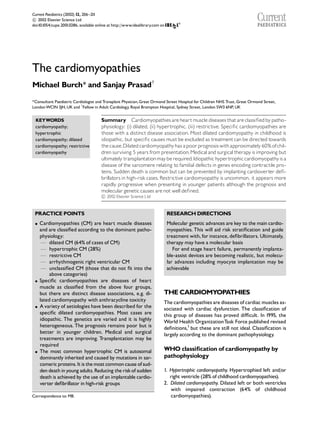
Call Girls Hosur Just Call 7001305949 Top Class Call Girl Service Available
The cardiomyopathies
1. The cardiomyopathies
Michael Burch* and Sanjay Prasadw
*Consultant Paediatric Cardiologist and Transplant Physician,Great Ormond Street Hospital for Children NHS Trust,Great Ormond Street,
LondonWC1N 3JH,UK and w
Fellow in Adult Cardiology, Royal Brompton Hospital, Sydney Street, London SW3 6NP,UK
KEYWORDS
cardiomyopathy;
hypertrophic
cardiomyopathy; dilated
cardiomyopathy; restrictive
cardiomyopathy
Summary Cardiomyopathies areheartmuscle diseasesthat are classi¢edbypatho-
physiology: (i) dilated, (ii) hypertrophic, (iii) restrictive. Speci¢c cardiomyopathies are
those with a distinct disease association. Most dilated cardiomyopathy in childhood is
idiopathic, but speci¢c causes must be excluded as treatmentcan be directed towards
thecause.Dilatedcardiomyopathyhasapoorprognosiswithapproximately 60% ofchil-
dren surviving 5 years frompresentation.Medical and surgicaltherapyisimproving but
ultimately transplantationmayberequired.Idiopathichypertrophiccardiomyopathyisa
disease of the sarcomere relating to familial defects in genes encoding contractile pro-
teins. Sudden death is common but can be prevented by implanting cardioverter de¢-
brillators in high-riskcases.Restrictive cardiomyopathyis uncommon, it appears more
rapidly progressive when presenting in younger patients although the prognosis and
moleculargenetic causes are not wellde¢ned.
c 2002 Elsevier Science Ltd
PRACTICE POINTS
K Cardiomyopathies (CM) are heart muscle diseases
and are classi¢ed according to the dominant patho-
physiology:
F dilated CM (64% of cases of CM)
F hypertrophic CM (28%)
F restrictive CM
F arrhythmogenic right ventricular CM
F unclassi¢ed CM (those that do not ¢t into the
above categories)
K Speci¢c cardiomyopathies are diseases of heart
muscle as classi¢ed from the above four groups,
but there are distinct disease associations, e.g. di-
lated cardiomyopathy with anthracycline toxicity
K Avariety of aetiologies have been described for the
speci¢c dilated cardiomyopathies. Most cases are
idiopathic. The genetics are varied and it is highly
heterogeneous. The prognosis remains poor but is
better in younger children. Medical and surgical
treatments are improving. Transplantation may be
required
K The most common hypertrophic CM is autosomal
dominantly inherited and caused by mutations in sar-
comericproteins.Itis themostcommoncause of sud-
den deathinyoung adults.Reducing theriskof sudden
death is achieved by the use of an implantable cardio-
verter de¢brillator in high-risk groups
RESEARCHDIRECTIONS
Molecular genetic advances are key to the main cardio-
myopathies. This will aid risk strati¢cation and guide
treatmentwith, for instance, de¢brillators. Ultimately,
therapy may have a molecular basis
For end stage heart failure, permanently implanta-
ble-assist devices are becoming realistic, but molecu-
lar advances including myocyte implantation may be
achievable
THE CARDIOMYOPATHIES
The cardiomyopathies are diseases of cardiac muscles as-
sociated with cardiac dysfunction. The classi¢cation of
this group of diseases has proved di⁄cult. In 1995, the
World Health OrganizationTask Force published revised
de¢nitions,1
but these are still not ideal. Classi¢cation is
largely according to the dominant pathophysiology.
WHO classi¢cation of cardiomyopathy by
pathophysiology
1. Hypertrophic cardiomyopathy. Hypertrophied left and/or
right ventricle (28% of childhood cardiomyopathies).
2. Dilated cardiomyopathy. Dilated left or both ventricles
with impaired contraction (64% of childhood
cardiomyopathies).Correspondence to: MB.
Current Paediatrics (2002) 12, 206^211
c 2002 Elsevier Science Ltd
doi:10.1054/cupe.2001.0286, available online at http://www.idealibrary.com on
2. 3. Restrictive cardiomyopathy. Restrictive ¢lling and
reduced diastolic volume of either or both ventricles
with normal or near normal systolic function and wall
thickness.
4. Arrythmogenicrightventriclecardiomyopathy.Progressive
¢bro fatty replacement of the right ventricular
myocardium later involving the left ventricle.
5. Unclassi¢edcardiomyopathies.
In the past, cardiomyopathies were de¢ned as being of
unknown cause and were, therefore, considered sepa-
rately from heartmuscleproblems causedbyknown dis-
eases. In recent years, with advances in molecular
genetics, the underlying disease processes are beginning
to be understood for many of the cardiomyopathies, so
that subdivisionsinto primary and secondarycardiomyo-
pathies is no longer relevant.
Speci¢c cardiomyopathies
Heart muscle problems associated with known diseases
are currently termed‘speci¢c cardiomyopathies’, and are
a subclassi¢cation of thepathophysiologicalgroup, i.e. di-
lated cardiomyopathy may be secondary to adriamycin
toxicity. When no association is known, the cardiomyo-
pathy may just be de¢ned by its pathophysiology (e.g.
dilated cardiomyopathy) or it could be termed idiopathic
dilated cardiomyopathyFalthough this term is not com-
monlyused.The groups of speci¢c cardiomyopathies and
the associated pathophysiological types are listed in
Table 1 as diseases of the myocardium associated with
cardiac dysfunction.
The ¢rst four of the speci¢c cardiomyopathies listed
in Table 1 are predominantly adult diseases. Ischaemic,
valvular and hypertensive cardiomyopathies have cardiac
disease out of proportion to the primary problem.
Peripartum cardiomyopathy is a mixedgroup of diseases
causing cardiac dysfunction in the perinatal period.
In£ammatory cardiomyopathy is myocarditis with car-
diac dysfunction. It is ‘dilated’ by pathophysiology. It may
be infectious, typically enteroviral (coxsackie) or adeno-
virus, but many other viruses including HIVand hepatitis
C and non-viral causes (bacteria, fungal, protozoal) are
known to occur. Fulminant myocarditis appears to have
a better prognosis than chronic in£ammation, and com-
plete recovery can occur. Histologically giant cell forma-
tion is associated with a poor prognosis. In general, the
managementis similar to idiopathic dilated cardiomyopa-
thy. Immune suppression and immunoglobulin therapy
await evaluation in randomized trials. In South America,
Chagas disease is a common cause.
Autoimmune causes of dilated cardiomyopathy are
well described and are seen with connective tissue dis-
eases, SLE, polyarteritis, rheumatoid, scleroderma and
dermatomyositis.Other systemic diseases thatcan cause
cardiomyopathy include sarcoidosis and leukaemia.
The muscular dystrophies are associated with cardiac
muscle disease, Duchenne being associated with hyper-
trophic changes initially, with dilated cardiomyopathy de-
veloping later. Becker cardiomyopathy is usually, but not
always, less severe than Duchenne. Some X-linked cases
of dilated cardiomyopathy without skeletal myopathy
have been shown to have de¢cient cardiac dystrophin.
Metabolic cardiomyopathy includes inborn errors of
metabolism and mitochondrial diseases. Mitochondrial
disease may be suspected when there is a maternal in-
heritance, epilepsy, familial diabetes, deafness and skele-
tal myopathy. Initially, hypertrophic changes may be seen
with poor contraction. Barth syndrome is a mitochon-
drial disease with dilated cardiomyopathy, it is X-linked
and there is cyclical neutropaenia.Improvement may oc-
cur with carnitine therapy (as with other mitochondrial
diseases).In¢ltration of the myocardium occurs in a vari-
ety of cardiomyopathies causing a range of pathophysiol-
ogies including hypertrophic, dilated and restricted.
Causesinclude Pompes disease (glycogen storage disease
II), which causes a severe hypertrophic cardiomyopathy
and is usually fatal in infancy. Fabrys, haemochromatosis
and amyloidosis are more severe in adult life.
Toxic reactions can cause dilated cardiomyopathy;
this can occur with anthracycline, radiation, alcohol and
cocaine abuse.
Dilated cardiomyopathy (Fig.1)
The speci¢c associateddiseases are discussed above, and
should always be sought. Most dilated cardiomyopathy
(DCM) in childhood is of unknown cause and this can be
frustrating when extensive investigations for associated
disease are negative. Paediatricians must be aware that
congenital heart disease, such as an anomalous coronary
artery, can present as a dilated cardiomyopathy.Thismay
be apparent on echocardiography but angiography is
sometimes needed.The aetiology of the idiopathic group
Table 1 Speci¢ccardiomyopathies*
Disease Type ofcardiomyopathy
Ischaemic CM DCM
Valvular CM DCM/HCM
Hypertensive CM HCM/RCM
Peripartum CM DCM
In£ammatory CM DCM
Metabolic DCM/HCM/RCM
Generalsystem disease DCM
Musculardystrophies DCM/HCM
Neuromusculardisorders HCM/DCM
Sensitive andtoxic reactions DCM
*CM, cardiomyopathy; D, dilated; H, hypertrophic; R,
restrictive.
THE CARDIOMYOPATHIES 207
3. is probably varied and viral myocarditis (acute and
chronic) and autoantibody disease may contribute. Spe-
ci¢c abnormalities of the myocyte cytoskeleton have
been detected and a widevariety of inheritance patterns
recordedincludingrecessive, X-linkedanddominant (the
most common). There is an age-related penetrance,
which makes screening and counselling di⁄cult. The in-
heritance has been described as a molecular maze.2
Re-
cently, an abnormality in myosin has been described in
familial dilated cardiomyopathy.3
Treatment is essentially that of chronic heart failure.
Diuretics areused torelieve symptoms.Data from excel-
lent adult trials of medical therapy can be used to guide
paediatric practice, where smaller numbers make such
studies di⁄cult. In essence, there is now overwhelming
evidence in favour of the use of angiotensin converting
enzyme inhibitors such as enalapril and captopril.4
Aldosterone antagonists such as spironolactone are also
bene¢cial and beta-blockers too can be used e¡ectively,
particularly carvedilol.5
The prognosis of DCM in
childhood is poor with 5-year survival of 60%,6
with
many deaths occurring shortly after presentation.
The outlook appears better in children under 2 years
of age. Surgical techniques such as mitral valve surgery
and ventricular reduction have been undertaken in
adults, but are used less widely in children.Left ventricu-
lar assist devices may be required for intractable heart
failure.7
Ultimately, cardiac transplantation may be
required.
Hypertrophiccardiomyopathy (Fig. 2)
Hypertrophic cardiomyopathy represents a heteroge-
nous group of disorders, and this diversityismore appar-
ent in childhood than at any other age (Table 2).
Familialhypertrophiccardiomyopathy
Hypertrophic cardiomyopathy is a primary disease of
cardiac muscle in the absence of valvar stenosis, hyper-
tension, or other disease processes. Inheritance is auto-
somal dominant with variable phenotypic expression.
Defects in genes encoding three contractile proteins
(cardiac troponinT, beta-myosin heavy chain, and alpha-
tropomyosin) can create the phenotypic expression.8,9
Ultimately, the diagnosis of familial hypertrophic cardio-
myopathy depends on molecular identi¢cation of the of-
fending gene or the abnormal gene product.
Histologically, there is myocyte disarray.The prevalence
is around 0.2% of the population.
In the UK, this disorder is a leading cause of sudden
death, particularly in otherwise healthy young persons
such as athletes. Familial hypertrophic cardiomyopathy
is characterized by myocardial hypertrophy and a wide
spectrum of symptoms, including dyspnoea, palpitations,
light-headedness, chest pain and syncope. Syncope oc-
cursin15^25% of adult subjects. Although syncopeisless
common in childhood, it is strongly associated with the
risk of sudden death. There is an annual death rate of
2^4% from sudden death, which can occur even in
asymptomatic individuals.
Electrocardiography results are abnormal in about
90% of patients and may show a wide variety of patterns.
Echocardiographic features of hypertrophic cardiomyo-
pathy have been well described. Mild or marked left,
right or biventricular hypertrophy can be detected by
echocardiography.The distribution of hypertrophy in hy-
pertrophic cardiomyopathy is characteristically asym-
metrical, less commonly it is symmetrical or apical.The
anatomical pattern has not proved to be predictive of
outcome but is a primary determinant of out£ow ob-
struction and is an important factor in surgical planning.
Figure 1 MRI scan of a patient with dilated cardiomyopathy. Left-hand ¢gure shows the heart in end-diastole; right-hand ¢gure
shows the heartin end-systole.Theleft ventricleisgrosslydilated and functionis severelyimpaired.
208 CURRENT PAEDIATRICS
4. Other echocardiographic ¢ndingsinclude dynamic mitral
regurgitation and left ventricular out£ow obstruction.
Out£ow obstruction is present in less than half of the
patients with familial hypertrophic cardiomyopathy and
is not predictive of outcome, with symptomatic patients
without obstruction faring more poorly than those who
have gradients. The magnitude of out£ow obstruction
appears unrelated to the occurrence of ventricular
tachycardia or risk of sudden death.
High-grade arrythmias are elicited in some patients
and have a negative prognostic implication. A hypoten-
sive response to exercise appears to represent a risk for
sudden death but more de¢nitively, a normal exercise
blood pressure response identi¢es a low-risk cohort.
Primary histological abnormality of focal myocardial
disarray is not unique to familial hypertrophic
cardiomyopathy and cannot be reliably detected on
biopsy specimens.
Di¡erentiation between physiological hypertrophy
secondary to athletic participation and pathological hy-
pertrophy in familial hypertrophic cardiomyopathy is a
frequent and important problem in young adults. The
cardiac responses to chronic, intensive exercisehasbeen
well characterized and include dilation and hypertrophy
with preservation of myocardial contractility.The hyper-
trophic response is most intense in sports that elicit a
marked rise in blood pressure during exercise, such as
rowing, wrestling and power lifting. Wall thickness
413mm, is occasionally found in athletes, and the not
infrequent occurrence of mild left ventricular hypertro-
phy in patients with familial hypertrophic cardiomyopa-
thy result in a signi¢cant incidence of diagnostic
ambiguity.ECGhas notbeen particularlyhelpfulin di¡er-
entiation because of the frequent presence of ECG ab-
normalities in athletes. Echocardiographic and clinical
features that increase the probability of familial hyper-
trophic cardiomyopathies include:
(a) a family history of hypertrophic cardiomyopathy or
early sudden death
(b) signi¢cant regional di¡erences in hypertrophy
(c) diastolic dysfunction
(d) abnormal ultrasonic myocardial re£ectivity
(e) absence of deconditioning-induced regression of
hypertrophy, and
Figure 2 Cardiac MRI scan of a patient withhypertrophiccardiomyopathy.Topleft ¢gure shows a four-chamber view demonstrat-
ing gross hypertrophy of the left ventricular wall.Top right ¢gure shows the left ventricular out£ow tract.The bottom two show a
short-axis view ofthethickenedleft ventriclein end-diastole and end-systole, respectively.
Table 2 Causes of hypertrophy
1. Hypertension
2. Congenitalheartdisease
3. Infantofdiabetic mother
4. Drugs, e.g. prenatal and postnatal corticosteroids,
tacrolimus, anabolic steroids
5. Metabolicdisease, e.g.GSDII,III, and IV,Fabrys,Icelldisease,
mucopolysaccharidosis, carnitine de¢ciency
6. SyndromesFNoonan, Leopard, Friedreich’s ataxia,
Beckwith^Weidemann,Costello
7. Familialhypertrophiccardiomyopathy
THE CARDIOMYOPATHIES 209
5. (f) abnormalities in coronary £ow reserve.
Ultimately, di¡erentiation by available techniques is sim-
ply not possible in some subjects.
In infants, restrictive symptoms predominate. High
dose beta-blockers may be helpful; disopyramide has
beenused to reduce the out£owgradient. Surgery, asyn-
chronous dual chamber pacing, and non-surgical septal
ablation are all treatment options where pharmacologi-
cal agents have been unsuccessful. Surgical or pharmaco-
logical reduction in the out£ow gradient in symptomatic
patients is usually associated with a reduction in symp-
toms, although the incidence of sudden death is not im-
proved. In general, dynamic out£ow obstruction is not a
negative prognostic factor, and interventions aimed at
reducing the gradient are justi¢ed only in as much as
symptomatic bene¢t can be anticipated.
Ventricular tachycardia or ¢brillation is probably the
mechanism of sudden death in hypertrophic cardiomyo-
pathy. The implantable de¢brillator is highly e¡ective in
terminating malignant ventricular arrhythmias in these
patients and shouldbe o¡ered to patientsin the high-risk
category for primary and secondary prevention of sud-
den death.10
Avoidance of strenuous exercise is generally
recommended for patients with familial hypertrophic
cardiomyopathy.
Major adverse risk factors include a family history of
sudden death, resuscitated cardiac arrest, exercise-
induced hypotension, syncope and symptomatic non-
sustained ventricular tachycardia on Holter recording.
Additionally, the extent of hypertrophy may be prognos-
tic.Patients free of allrisk factors are considered to be at
low risk, and interventions (other than for symptoms
such as chest pain or exercise intolerance) are not indi-
cated. With two or more risk factors or with syncope
alone, riskis consideredhigh and aggressivemanagement
such as with an implantable cardioverter-de¢brillator is
recommended.No consensus has been reached on man-
agement of intermediate-risk patients. Additional nega-
tive prognostic factors such as evidence of ischaemia on
exercise thallium testing, marked QT dispersion, and
myocardialbridgingcan alsobeusefulinmanagementde-
cisions for these patients.Genotyping may help more ac-
curate risk strati¢cation and guidance of treatment.11
Systolic function is nearly always normal or hyperdy-
namic. Sudden death in patientsreferred to tertiary care
centres is seen annually in 3^5% of adults and 6^8% of
children. Recent population studies indicate a much low-
er annual mortality (0.1^1%), which indicates a major
referral bias in these statistics.12
Restrictive cardiomyopathy (Fig. 3)
This is the least common form of cardiomyopathy and
is unusual among children, where causes include
some forms of storage disease.13
Clinical features are
Figure 3 Transoesophagealechocardiogramofapatientwith
advanced cardiac in¢ltrative disease (top ¢gure) shows thick-
ened myocardial walls and restrictive physiologic features with
markedly decreased ratio of pulmonary venous systolic-to-
diastolic £ow (middle ¢gure) and shortened deceleration time
(100 ms) oftransmitralin£ow E-wave velocity (bottom ¢gure).
210 CURRENT PAEDIATRICS
6. comparable to those in adults, with normal ventricular
size and function, severe elevationin diastolic ¢llingpres-
sure and distinct atrial dilatation.14
Unlike adults, paedia-
tric cases have been almost consistently idiopathic
despite tissue analysisin nearly all, although several cases
were familial. Di¡erentiation from many of the second-
ary causes, such as myocardial non-compaction (persis-
tence of embryonic or ‘spongy’ myocardium), can be
made on morphological criteria. Endomyocardial biopsy
is sometimesundertaken to exclude anypotentially trea-
table disorder. A striking feature in children is the poor
prognosis, with a 2-year survival rate of about 50%.15
Survival, therefore, appears to be even more limited
than has been described in adults.Younger patients with
respiratory symptoms, thromboembolism, increased
cardiothoracic ratio on chestradiogram or patients with
endocardial ¢broelastosis appear to have a worse
prognosis.
Anticoagulation is recommended because a 25% inci-
dence of thromboembolism has been seen in children.
Therapy is otherwise non-speci¢c and usually is of very
limitedbene¢t.The onset of irreversible elevation in pul-
monary vascular resistance can occur within 1^4 years
in these patients, and early cardiac transplantation is
therefore recommended to avoid the need for heart
and lung transplantation.
Right ventriculardysplasia
Right ventricular dysplasia is an idiopathic cardiomyopa-
thy associated with sudden cardiac death. It is of unclear
aetiology but thought to be an autosomal dominant dis-
order with variable expression and penetrance.This car-
diomyopathy mainly a¡ects the right ventricle although
the left ventricle may also be a¡ected. Histologically, it
is characterized by a lipomatous or ¢brolipomatous
transformation of the right ventricular myocardium.The
presence of adipose tissue together with ¢brosis and
myocyte hypertrophy in young patients strongly sug-
gests right ventricular dysplasia.Patients commonly pre-
sentwith asymptomatic cardiomegaly (10%) orrecurrent
ventricular arrhythmias of leftbundle branch block mor-
phology. It has been described as a cause of ventricular
tachycardia in children with apparently normal hearts.
A family history of cardiomyopathy, or sudden death in a
close relative also can be a clue to the diagnosis.16,17
On ECG, typically there is aT-wave inversion in right
precordial leads and localized prolongation of QRS com-
plex in right precordial leads.Ventricular tachycardia and
frequentventricular extrasystolesmaybe seen.On echo,
or better MRI, there may be cardiomegaly with a dilated
impaired right ventricle.
REFERENCES
1. Richardson P, McKenna W, Bristow M et al. Report of the World
Health Organisation/International Society and Federation of Car-
diology Task Force on the de¢nition and classi¢cation of cardio-
myopathies.Circulation1996; 93(5): 841.
2. Komajda M.Genetics of dilated cardiomyopath: a molecular maze?
Heart 2000; 84(5): 463^464.
3. Kamisago M, Sharma S D, De Palma S R et al. Mutations in sarco-
mere protein genes as a cause of dilated cardiomyopathy. N Engl J
Med 2000; 343(23):1688^1696.
4. The SOLVD Investigators.N Engl J Med1991; 325(5): 293^302.
5. Packer M,Coats A S, Fowler M R et al. E¡ect of Carvedilol on sur-
vival in severe chronic heart failure. N Engl J Med 2001; 344:1651^
1658.
6. Burch M, Siddiqi S A,Celermajer D E et al.Dilatedcardiomyopathy
in children: determinants of outcome.Br Heart J1994; 72(3): 246^
250.
7. Westaby S, Franklin O, Burch M. New developments in the treat-
ment of cardiac failure. Arch Dis Child1999; 81(3): 267^277.
8. Burch M, Blair E.The inheritance of hypertrophic cardiomyopathy.
Pediat Cardiol1999; 20(5): 313^316.
9. Marian A J. On genetic and phenotypic variability of hypertrophic
cardiomyopathy: nature versus nurture. J Am Coll Cardiol 2001;
38(2): 331^334.
10. Maron B J,Wing-Kuang Shen,Link M S et al.E⁄cacyof implantable
cardioverter-de¢brillators for the prevention of sudden death in
patients with hypertrophic cardiomyopathy. N Engl J Med 2000;
342: 365^373.
11. Havndrup O, Bundgaard H, Andersen P S, Larsen L A et al. The
Val606Met mutation in the cardiac beta-myosin heavy gene in pa-
tients with familial hypertrophic cardiomyopathy is associated
with a high risk of sudden death at a young age. Am J Cardiol 2001;
87(11):1315^1317.
12. Maron B J, Casey S A, Poliac L C et al. Clinical course of hyper-
trophic cardiomyopathy in a regional United States cohort. JAMA
1999; 281: 650.
13. Schutte D P, Essop M R.Clinical pro¢le and outcome of idiopathic
restrictive cardiomyopathy.Circulation 2001; 103(14): E83.
14. Hancock E W. Di¡erential diagnosis of restrictive cardiomyopathy
and constrictive pericarditis.Heart 2001; 86(3): 343^349.
15. Chen S C, Balfour I C, Jureidini S. Clinical spectrum of restrictive
cardiomyopathy in children. J Heart LungTransplant 2001; 20(1):
90^92.
16. Corrado D, Fontaine G, Marcus F L et al. Arrhythmogenic right
ventricular dysplasia/cardiomyopathy: need for an international
registry. Study Group on Arrhythmogenic Right Ventricular Dys-
plasia/Cardiomyopathy of the Woking Groups on Myocardial and
Pericardial Disease and Arrhythmias of the European Society Of
Cardiology and of the Scienti¢c Council on Cardiomyopathies of
the World Heart Federation.Circulation 2000; 101(11): E101^E106.
17. Oakley R M,Ooi O C, Bongso A et al.Myocyte transplantation for
myocardial repair: a few good cells can mend a broken heart. Ann
Thorac Surg 2001; 71(5):1724^1733.
THE CARDIOMYOPATHIES 211