Examination of the gingival unit and alveolar mucosa may be done second in an organized sequence of a comprehensive periodontal evaluation, See this chart which is POSTED with your other October handouts on the ADHP site. Learn the symbols here related to charting the tooth AND gingival feat- ures AND those pertain-ing to examination of the attachment apparatus (pocket depth, etc.) The methods of physical examination of the gingiva are mainly by (1) Inspection, (2) Palpation of the dried tissues. These are general manifestations and some are more (or less) pronounced in certain sites – i.e.gingiva. Features Of The Gingiva To Evaluate To Decide If It is Healthy Or Diseased. Color, Contour (Shape), Consistency, Size, Bleeding On Touch or BOP, Texture.Clinical Signs And Symptoms Of Gingivitis- Change in Color – Red Cyanotic, Increase in Size and Shape, Change in Consistency – Edematous (Soft) or Fibrotic (Firm), Bleeding, Exudate -- Gingival (Crevicular) Fluid, Pain. Clinical Signs And Symptoms Of Gingivitis- Change in Color : Pink, Red Cyanotic, Increase in Size and Shape, Change in Consistency – Edematous (Soft), Fibrotic (Hard). Gingival Bleeding- “Spontaneous” Bleeding: Bleeds from slight con- tact by probe or air to external surface of gin- giva (not in sulcus).
Call Girls Service In Shyam Nagar Whatsapp 8445551418 Independent Escort Service
Examination Of The Gingival Unit and Alveolar Mucosa
1. Examination Of The Gingival Unit and Alveolar Mucosa
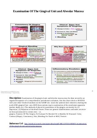
Description: Examination of the gingival unit and alveolar mucosa may be done second in an
organized sequence of a comprehensive periodontal evaluation, See this chart which is POSTED
with your other October handouts on the ADHP site. Learn the symbols here related to charting the
tooth AND gingival feat- ures AND those pertain-ing to examination of the attachment apparatus
(pocket depth, etc.) The methods of physical examination of the gingiva are mainly by (1)
Inspection, (2) Palpation of the dried tissues. These are general manifestations and some are more
(or less) pronounced in certain sites – i.e.
gingiva. Features Of The Gingiva To Evaluate To Decide If It is Healthy Or Diseased. Color,
Contour (Shape), Consistency, Size, Bleeding On Touch or BOP, Texture.
Reference Url : http://medical.wesrch.com/paper-details/pdf-ME1LYYEUOHRIY-examination-
of-the-gingival-unit-and-alveolar-mucosa