SlideShare a Scribd company logo
1 of 8
Presented by: Dave Jay S. Manriquez RN.
Veins
   • are thin-walled vessels that transport deoxygenated blood from the capillaries
      back to the right side of the heart

3 Layers – intima, media, adventitia

   •   there is little smooth muscle & connective tissue Ú makes the veins more
       distensible Ú they accumulate large volumes of blood
   •   Major veins, particularly in the lower extremities, have one-way valves ---allow
       blood flow against gravity
   •   Valves allow blood to be pumped back to the heart but prevent it from draining
       back into the periphery

Vein Disorders
      Venous Thrombosis (Superficial and Deep Vein Thrombosis), Thrombophlebitis,
        Phlebothrombosis – the above terms do not necessarily represent an identical
        pathology, for clinical purposes they are often used interchangeably.
Cause of Venous thrombosis remains unclear, three antecedents factors are believed to
play a significant role in its development: stasis of blood, injury to the vessel wall, and
altered blood coagulation.
Thrombophlebitis – is inflammation of the walls of the veins, often accompanied by the
formation of a clot. When a clot develops initially in the veins as a result of stasis or
hypercoagulability , but without inflammation, the process is referred to as
Phlebothrombosis.
Venous thrombosis can occur in any vein but is most frequent in the veins of the lower
extremities. Both superficial and deep veins of the legs may be affected. Of the superficial
veins, the saphenous vein is most frequently affected. Of the deep leg veins, the
iliofemoral, popliteal, and small calf veins are most often involved.

Chronic Venous Insufficiency - Venous insufficiency is a disease state resulting from the
obstruction or reflux of venous valves in the legs. Both superficial and deep leg veins can
be involved. The resulting venous hypertension can occur whenever there has been a
prolonged increase in venous pressure, such as occurs with deep venous thrombosis. The
walls of veins are thinner and more elastic than walls of arteries, they distended readily
when venous pressure is consistently high. In this state, leaflets of the venous valves are
stretched and prevented from closing completely, thereby allowing a backflow or reflux
of blood in the veins.

When a deep veins in the legs have incompetent valves after a thrombus, postphlebitic
syndrome may develop. This result in edema, altered pigmentation, pain, stasis
dermatitis, and stasis ulceration.

    Varicose Veins - abnormally dilated, tortuous, superficial            veins cause by
     incompetent venous valves.
Venous Disorders
    alteration in the transport/flow of blood from the capillary back to the heart
    changes in smooth muscle and connective tissue make the veins less distensible
      with limited recoil capacity
    valves may malfunction, causing backflow of blood

    Virchow’s triad: blood stasis, vessel wall injury, and altered blood coagulation

Thrombophlebitis
    inflammation of the veins caused by thrombus or blood clot
Factors assoc. with the devt. of Thrombophlebitis
    venous stasis – occurs when blood flow is retarded, such as heart failure and
       shock; when veins are dilated, such as after drug therapy; and when skeletal
       muscle contraction is reduced, as with immobility, extremity paralysis, or
       anesthesia.
    damage to the vessel wall – disruption of the intimal lining of blood vessel creates
       a site for clot formation, such as after a fracture or dislocation, diseases of the
       veins, and chemical irritation of the vein from intravenous drugs or solutions.
    hypercoagulability of the blood – oral contraceptive use
    common to hospitalized pts. , undergone major surgery (pelvic or hip surgery),
       MI
Pathophysiology
    develops in both the deep and superficial veins of the lower extremity
    deep veins – femoral, popliteal, small calf veins
    superficial veins – saphenous vein
    Thrombus – form in the veins from accumulation of platelets, fibrin, WBC and
       RBC

Risk Factors for Thrombophlebitis
    Bed Rest
    General Surgery
    Leg Trauma
    Previous Venous Insufficiency
    Obesity
    Oral Contraceptives
    Malignancy

Deep Vein Thrombosis (DVT)
     tends to occur at bifurcations of the deep veins, which are sites of turbulent blood
       flow
     a major risk during the acute phase of thrombophlebitis is dislodgment of the
       thrombus Ú embolus
     pulmonary embolus – is a serious complication arising from DVT of the lower
       extremities
Clinical Manifestations:
     pain and edema of extremity – obstruction of venous flow
 Û circumference of the thigh or calf
     (+) Homan’s sign – dorsiflexion of the foot produces calf pain
     Do not check for the Homan’s sign if DVT is already known to be present Ú Û
       risk of embolus formation
     * if superficial veins are affected - signs of inflammation may be noted – redness,
       warmth, tenderness along the course of the vein, the veins feel hard and thready &
       sensitive to pressure
The risk of dislodgement and embolization of superficial venous thrombi is very low
because the majority of them undergo spontaneous lysis, thus this condition can be
treated at home with rest, extremity elevation, analgesics, and possibly anti-
inflammatory agents.

Diagnostic Evaluation
Noninvasive Techniques: - rely on the thrombus to create abnormalities of venous flow.
     Doppler Ultrasonography – use of a Doppler probe placed over veins that are
        obstructed. Nonexpensive, portable, simple, rapid, and noninvasive.
     Duplex Venous Imaging – able to obtain anatomic information, as well as to
        assess physiologic parameters.
     Impedance Plethysmography – is used to measure changes in venous vlume, a
        blood pressure cuff is applied to patient thigh inflated enough to impede venous
        flow (50-60 mmHg), calf electrodes are used to measure electrical resistance that
        results from venous volume changes.
Invasive Techniques: - rely on the injection of contrast media into the venous system,
which then bind with structural elements of the thrombus.
I-labeled Fibrinogen Scanning - a sensitive method for early detection of venous
thrombosis. The test relies on the fact that radioactive fibrinogen, when injected
intravenously, will concentrate in the forming clot. The level o radioactivity can then be
serially measured by an external counter, and the progression of the clot can be
monitored.
Contrast Phlebography - involves the injection of radiographic contrast media into the
venous system through a dorsal foot vein.

Medical Management
Superficial thrombophlebitis
    bed rest with legs elevated
    apply moist heat
    NSAID’s ( Non – steroidal anti-inflammatory drugs) - aspirin
Deep vein thrombosis
    requires hospitalization
    bed rest w/ legs elevated to 15-20 degrees above heart level ( knees slightly
       flexed, trunk horizontal, head may be raised) to promote venous return and help
       prevent further emboli and prevent edema
    application of warm moist heat to reduce pain, promotes venous return
    elastic stocking or bandage
    anticoagulants, initially with IV heparin then coumadin
    fibrinolytic to resolve the thrombus
 vasodilator if needed to control vessel spasm and improve circulation
Nursing Intervention
Preventive care
    prevent long periods of standing or sitting that impair venous return
    elevate legs when sitting, dorsiflex feet at regular intervals to prevent venous
       pooling
    if edema occurs, elevate above heart level
    regular exercise program to promote circulation
    avoid crossing legs at the knees
    avoid wearing constrictive clothing such as tight bands around socks or garters
    use elastic stocking on affected leg
    do leg exercises during periods of enforced immobility such as after surgery

Nursing Assessment - is invaluable in detecting early signs of venous disorders of the
lower extremities. Patients with history of varicose veins, hypercoagulation, neoplastic
disease, cardiovascular disease, or recent major surgery or injury, and the obese, the
elderly, and women taking oral contraceptives are in the high risk group.
     characteristic of the pain
     onset & duration of symptoms
     history of thrombophlebitis or venous disorders
     color & temp. of extremity
     edema of calf of thigh - use a tape measure, measure both legs for comparison
     Identify areas of tenderness and any thrombosis
Surgery
             if the thrombus is recurrent and extensive or if the pt. is at high risk for
                pulmonary embolism - . Surgery for deep vein thrombosis is necessary
                when: 1. anticoagulant or thrombolytic therapy is contraindicated. 2. the
                danger of pulmonary embolism is extreme and 3. the venous drainage is
                so severely compromised that permanent extremity damage will probably
                result.
     Thrombectomy – incising the common femoral vein in the groin and extracting
        the clots
     Vena caval interruption – transvenous placement of a grid or umbrella filter in the
        vena cava to block the passage of emboli

Nursing Management
Acute care
    explain purpose of bed rest and leg elevation
    use elastic stockings
    monitor pt. on anticoagulant & fibrinolytic therapy for signs of bleeding
    monitor for signs of pulmonary embolism – sudden onset of chest pain, dyspnea,
       rapid breathing, tachycardia
Nsg. intervention often surgery of vena caval interruption
    assess insertion site – bleeding, hematoma, apply pressure over site and inform
       physician
 keep pt. on bed rest for 1st 24 hrs. then encourage ROM exercises to promote
     venous return
    assist pt. in ambulation when permitted, elevate legs when sitting
    keep elastic bandage
    avoid rubbing or massaging the affected extremity
    give analgesic and anti-inflammatory agents to promote comfort

Anticoagulant Therapy for Thromboembolism:
- is the administration of a medication to delay the clotting time of blood, to prevent the
formation of a thrombus in postoperative patients, and to forestall the extension of a
thrombus once it has formed. Anticoagulants cannot dissolve a thrombus that has already
formed.

Administration:
- Heparin is administered using a continuous pump infusion. To promptly reverse the
effects of heparin , the physician may prescribe intravenous injections of protamine
sulfate. For Coumadin it is Vitamin K.
- Intermittent intravenous injection is another means of administering heparin, in this
instance a dilute aqueous solution given every 4 hours.
- Oral anticougulants, such as Coumadin, are monitored by the prothrombin time.
Because Coumadin has a lag period of 3 to 5 days, it is usually administered in
conjunction with heparin until desired anticoagulation has been achieved.

Precaution and Nursing Assessment:
- The principal complication of anticoagulant therapy is the occurrence of spontaneous
bleeding anywhere in the body. A further possible complication of heparin therapy is that
of Heparin-induced thrombocytopenia which generally occurs 7 to 10 days after the
treatment has been started. Oral anticoagulants interact with many other medications. It
is advisory to study drug interactions for patients taking specific oral coagulants.

Patient Education About Oral Anticogulants:
- The patient should be informed about the medication, its purpose, and the need to take
the correct amount at the specific times prescribed, and should be aware that blood tests
are scheduled periodically to determine whether a change in medication dosage is
required.

Chronic Venous Insufficiency
   Results from obstruction of venous valves in legs or reflux of blood back through
      valves
   Venous ulceration is serious complication – this is a result of inadequate
      exchange of oxygen and other nutrients in the tissue, when the cellular
      metabolism cannot maintain energy balance, cell death (necrosis) results.
   Pharmacological therapy is antibiotics for infections
   Debridement to promote healing
   Topical Therapy may be used with cleansing and debridement
Management and Patient Education – management of the patient with venous
insufficiency is directed at reducing venous stasis and preventing ulcerations. Measures
that increases venous blood flow are antigravity activities and compression of superficial
veins with elastic stockings.

Elevation of the legs decreases edema, promote venous return. Elevations should be
performed frequently throughout the day (at least 30 minutes every 2 hours). At night, the
patient should sleep with the foot of the bed elevated for 6 inches. Prolonged sitting or
standing still detrimental, but walking should be encouraged. When sitting, patient
should avoid placing pressure on the popliteal spaces, such as occurs in leg crossing, or
sitting with the legs dangling over the side of the bed. Wearing of constricted garments
are also contraindicated.

Elastic stockings of the legs reduces pooling of venous blood and enhances venous return
to the heart. Prevent twisting of stockings causing tourniquet effect cause this will
worsens venous pooling. Extremities with venous insufficiency are conscientiously
protected from trauma. The skin is kept clean, dry, and soft. Signs of ulceration are
immediately reported to the nurse or physician for treatment and follow-up.

Varicose Veins
    are abnormally dilated veins with incompetent valves, occurring most often in the
       lower extremities, the saphenous veins, or the lower trunk however, it can occur
       elsewhere in the body. Affect1 of 5 persons in the world. This condition is most
       common in women and in persons in occupations requiring prolonged standing
       such as salespeople, barbers, beauticians, elevator operators, nurses and dentist.
       A hereditary weakness of the vein wall may contribute to the development of
       varicosities, and it is not uncommon to see this condition occur in several
       members of the same family.
   • usually affected are woman 30-50 years old.
Causes:
           – congenital absence of a valve
           – incompetent valves due to external pressure on the veins from pregnancy,
              ascites or abdominal tumors
           – sustained Û in venous pressure due to CHF, cirrhosis
Prevention
           – wear elastic stockings during activities that require long standing or when
              pregnant
           – moderate exercise, elevation of legs

Pathophysiology
     the great and small saphenous veins are most often involved
weakening of the vein wall does not withstand normal pressureäveins dilate , pooling of
bloodävalves become stretched and incompetentämore accumulation of blood in the
veins

Clinical Manifestations
 Primary varicosities – gradual onset and affect superficial veins, appearance of
       dark tortuous veins. Affected persons may have no symptoms, but cosmetically the
       appearance of the dilated vein is unappealing.
   S/sx – dull aches, muscle cramps, pressure, heaviness or fatigue arising from reduced
blood flow to the tissues
     Secondary Varicosities – affect the deep veins
            occur due to chronic venous insufficiency or venous thrombosis
  S/sx – edema, pain, changes in skin color, ulcerations may occur from venous stasis

Diagnostic Evaluation
A. Bordie-Trendelenburg test
    assess competency of venous valves through measurement of venous filling time
    the pt. lies down with the affected leg raised to allow for venous emptying
    a tourniquet is then applied above the knee and the pt. stands. The direction and
       filling time are recorded both before & after the tourniquet is removed
    * Incompetent valves are evident when the veins fill rapidly from backward blood
       flow

B. Perthes’ test - is a diagnostic procedure that easily indicates whether the deeper
venous system and communicating veins are competent. A tourniquet is applied just
below the knee and the patient is asked to walk. If the varicose veins disappear, the deep
system and communicating vessels are competent. If the vessels do not empty and become
distended on walking, incompetency or obstruction is inferred.

C. Additional tests for the presence of Varicose Veins are the Doppler flow meter- detect
retrograde flow of blood in superficial veins with incompetent valves after compression
of the leg proximally., Phlebography – the injection of radiographic contrast media into
the leg veins so that vein anatomy can be visualized during various leg movements. , and
Plethysmography – allows measurement of changes in venous blood volume..

Surgical Intervention
     indicated or done for prevention or relief of edema, for recurrent leg ulcers or pain
        or for cosmetic purposes
Vein ligation and stripping
     the great sapheneous vein is ligated (tied) close to the femoral junction
     the veins are stripped out through small incisions at the groin, above & below the
        knee and at the ankles.
     sterile dressing are placed over the incisions and an elastic bandage extending
        from the foot to the groin is firmly applied
DIAGRAM:
Ligation and stripping of the great and the small saphenous veins. (A) The tributaries of
the saphenous vein have been ligated, and the saphenous vein has been ligated at the
saphenofemoral junction . (B) Vein stripper has been inserted from the ankle superiorly
to the groin. The vein is stripped from above to downward. A number of alternate
incisions may be needed to remove separate varicosities masses. (C) The small
saphenous vein is stripped from its junction with the popliteal vein to a point posterior to
the lateral malleolus.

Sclerotherapy – an irritating chemical (Sotradecol) is injected into the vein, which
irritates venous endothelium and produces localized phlebitis and fibrosis, thereby
obliterating the lumen. This treatment may be performed alone for small varicosities or
may follow vein ligation and stripping. Sclerosing is a palliative not a curative treatment.

Nursing care after vein ligation & stripping
    Monitor for signs of bleeding, esp. on 1st post-op day
            if there is bleeding, elevate the leg, apply pressure over the wound and
              notify the surgeon
    Keep pt. flat on bed for first 4 hrs. after surgery, elevate leg to promote venous
       return when lying or sitting
    Medicate 30 mins. before ambulation and assist patient
    Keep elastic bandage snug and intact, do not remove bandage

More Related Content

What's hot (20)

varicose vein
varicose veinvaricose vein
varicose vein
 
Peptic ulcer.pptx
Peptic ulcer.pptxPeptic ulcer.pptx
Peptic ulcer.pptx
 
Varicose vein
Varicose  veinVaricose  vein
Varicose vein
 
Varicose vein AB
Varicose vein ABVaricose vein AB
Varicose vein AB
 
Aneurysm
Aneurysm Aneurysm
Aneurysm
 
DVT ppt.pptx
DVT ppt.pptxDVT ppt.pptx
DVT ppt.pptx
 
Portal Hypertension Mechanisms Pathophysiology by Dr. Aryan
Portal Hypertension Mechanisms Pathophysiology by Dr. AryanPortal Hypertension Mechanisms Pathophysiology by Dr. Aryan
Portal Hypertension Mechanisms Pathophysiology by Dr. Aryan
 
Pulmonary embolism (PE)
Pulmonary embolism (PE)Pulmonary embolism (PE)
Pulmonary embolism (PE)
 
varicose veins
varicose veinsvaricose veins
varicose veins
 
Venous Insufficiency
Venous InsufficiencyVenous Insufficiency
Venous Insufficiency
 
Varicose vein
Varicose veinVaricose vein
Varicose vein
 
Surgery 6th year, Tutorial (Dr. Aram Baram)
Surgery 6th year, Tutorial (Dr. Aram Baram)Surgery 6th year, Tutorial (Dr. Aram Baram)
Surgery 6th year, Tutorial (Dr. Aram Baram)
 
What is Phlebitis and How is it Treated?
What is Phlebitis and How is it Treated?What is Phlebitis and How is it Treated?
What is Phlebitis and How is it Treated?
 
Varicose veins and management 1
Varicose veins and management 1Varicose veins and management 1
Varicose veins and management 1
 
Slide for raynaud's disease
Slide for raynaud's diseaseSlide for raynaud's disease
Slide for raynaud's disease
 
Varicose veins
Varicose  veinsVaricose  veins
Varicose veins
 
Inguinal herniorrhaphy
Inguinal herniorrhaphyInguinal herniorrhaphy
Inguinal herniorrhaphy
 
L acute appendicitis
L acute appendicitisL acute appendicitis
L acute appendicitis
 
Varicose veins
Varicose veinsVaricose veins
Varicose veins
 
Dvt
Dvt Dvt
Dvt
 

Similar to Vein Disorder

Vein Disorders
Vein DisordersVein Disorders
Vein DisordersJack Frost
 
Nursing Care of Clients with Peripheral Vascular Disorders Part 3 of 3
Nursing Care of Clients with Peripheral Vascular Disorders Part 3 of 3 Nursing Care of Clients with Peripheral Vascular Disorders Part 3 of 3
Nursing Care of Clients with Peripheral Vascular Disorders Part 3 of 3 Carmela Domocmat
 
Vein Disorders
Vein DisordersVein Disorders
Vein DisordersJack Frost
 
DEEP VEIN THROMBOSIS (1) final.pptx
DEEP VEIN THROMBOSIS (1) final.pptxDEEP VEIN THROMBOSIS (1) final.pptx
DEEP VEIN THROMBOSIS (1) final.pptxAbhiL10
 
Fwd: Bambury lecture on venous and lymphatic disorders of the limb
Fwd: Bambury lecture on venous and lymphatic disorders of the limbFwd: Bambury lecture on venous and lymphatic disorders of the limb
Fwd: Bambury lecture on venous and lymphatic disorders of the limbJeku Jacob
 
Dvt Deep Venous Thrombosis
Dvt Deep Venous ThrombosisDvt Deep Venous Thrombosis
Dvt Deep Venous ThrombosisFazal Hussain
 
Venous drainage of lower limb ppt
Venous drainage of lower limb pptVenous drainage of lower limb ppt
Venous drainage of lower limb pptAditi Ujjawal
 
VARICOSE VEINS & DEEP VENOUS THROMBOSIS.pptx
VARICOSE VEINS & DEEP VENOUS THROMBOSIS.pptxVARICOSE VEINS & DEEP VENOUS THROMBOSIS.pptx
VARICOSE VEINS & DEEP VENOUS THROMBOSIS.pptxShahidHussain969204
 
Varicose veins and management 1
Varicose veins and management 1Varicose veins and management 1
Varicose veins and management 1sangu prashanth
 
Management of varicose veins RRT
Management of varicose veins RRTManagement of varicose veins RRT
Management of varicose veins RRTRanjith Thampi
 
8-Vein disorder.pptx
8-Vein disorder.pptx8-Vein disorder.pptx
8-Vein disorder.pptxssuser188360
 
dvt-131104063505-phpapp02.ppt
dvt-131104063505-phpapp02.pptdvt-131104063505-phpapp02.ppt
dvt-131104063505-phpapp02.pptafzal mohd
 

Similar to Vein Disorder (20)

D V T
D V TD V T
D V T
 
Vein Disorders
Vein DisordersVein Disorders
Vein Disorders
 
Nursing Care of Clients with Peripheral Vascular Disorders Part 3 of 3
Nursing Care of Clients with Peripheral Vascular Disorders Part 3 of 3 Nursing Care of Clients with Peripheral Vascular Disorders Part 3 of 3
Nursing Care of Clients with Peripheral Vascular Disorders Part 3 of 3
 
clinical aspects of vein
clinical aspects of veinclinical aspects of vein
clinical aspects of vein
 
Vein Disorders
Vein DisordersVein Disorders
Vein Disorders
 
deep vein thrombosis
deep vein thrombosisdeep vein thrombosis
deep vein thrombosis
 
Bindhya dvt
Bindhya dvtBindhya dvt
Bindhya dvt
 
DEEP VEIN THROMBOSIS (1) final.pptx
DEEP VEIN THROMBOSIS (1) final.pptxDEEP VEIN THROMBOSIS (1) final.pptx
DEEP VEIN THROMBOSIS (1) final.pptx
 
Deep Vein Thrombosis
Deep Vein ThrombosisDeep Vein Thrombosis
Deep Vein Thrombosis
 
VENOUS DISORDERS (thrombopheblitis)
VENOUS DISORDERS (thrombopheblitis)VENOUS DISORDERS (thrombopheblitis)
VENOUS DISORDERS (thrombopheblitis)
 
Fwd: Bambury lecture on venous and lymphatic disorders of the limb
Fwd: Bambury lecture on venous and lymphatic disorders of the limbFwd: Bambury lecture on venous and lymphatic disorders of the limb
Fwd: Bambury lecture on venous and lymphatic disorders of the limb
 
Varicose Vein
Varicose VeinVaricose Vein
Varicose Vein
 
Dvt Deep Venous Thrombosis
Dvt Deep Venous ThrombosisDvt Deep Venous Thrombosis
Dvt Deep Venous Thrombosis
 
Venous drainage of lower limb ppt
Venous drainage of lower limb pptVenous drainage of lower limb ppt
Venous drainage of lower limb ppt
 
Dvt
DvtDvt
Dvt
 
VARICOSE VEINS & DEEP VENOUS THROMBOSIS.pptx
VARICOSE VEINS & DEEP VENOUS THROMBOSIS.pptxVARICOSE VEINS & DEEP VENOUS THROMBOSIS.pptx
VARICOSE VEINS & DEEP VENOUS THROMBOSIS.pptx
 
Varicose veins and management 1
Varicose veins and management 1Varicose veins and management 1
Varicose veins and management 1
 
Management of varicose veins RRT
Management of varicose veins RRTManagement of varicose veins RRT
Management of varicose veins RRT
 
8-Vein disorder.pptx
8-Vein disorder.pptx8-Vein disorder.pptx
8-Vein disorder.pptx
 
dvt-131104063505-phpapp02.ppt
dvt-131104063505-phpapp02.pptdvt-131104063505-phpapp02.ppt
dvt-131104063505-phpapp02.ppt
 

More from Jack Frost

Other study questions - Related to Canadian Citizenship Exam
Other study questions - Related to Canadian Citizenship ExamOther study questions - Related to Canadian Citizenship Exam
Other study questions - Related to Canadian Citizenship ExamJack Frost
 
Important Names And Dates for Canadian Citizenship Exam
Important Names And Dates for Canadian Citizenship ExamImportant Names And Dates for Canadian Citizenship Exam
Important Names And Dates for Canadian Citizenship ExamJack Frost
 
90 ecom challenge
90 ecom challenge90 ecom challenge
90 ecom challengeJack Frost
 
Preceptorship meds
Preceptorship medsPreceptorship meds
Preceptorship medsJack Frost
 
Research for queens park common cases
Research for queens park common casesResearch for queens park common cases
Research for queens park common casesJack Frost
 
Diagnosis and medications research
Diagnosis and medications researchDiagnosis and medications research
Diagnosis and medications researchJack Frost
 
Jeopardy Game
Jeopardy Game Jeopardy Game
Jeopardy Game Jack Frost
 
Jeopardy 2016 - Handout
Jeopardy 2016 - HandoutJeopardy 2016 - Handout
Jeopardy 2016 - HandoutJack Frost
 
Cheat sheet - Plan of Care
Cheat sheet - Plan of CareCheat sheet - Plan of Care
Cheat sheet - Plan of CareJack Frost
 
Professional communication 4 simulation
Professional communication 4   simulationProfessional communication 4   simulation
Professional communication 4 simulationJack Frost
 
L 4 sims prep student 2016
L 4 sims prep student 2016L 4 sims prep student 2016
L 4 sims prep student 2016Jack Frost
 
Weekly reflections 2 and 3
Weekly reflections 2 and 3Weekly reflections 2 and 3
Weekly reflections 2 and 3Jack Frost
 
Horizontal violence prof comm4
Horizontal violence prof comm4Horizontal violence prof comm4
Horizontal violence prof comm4Jack Frost
 
Ethical dilemma class presentation
Ethical dilemma   class presentationEthical dilemma   class presentation
Ethical dilemma class presentationJack Frost
 
Teaching narcan injection
Teaching narcan injectionTeaching narcan injection
Teaching narcan injectionJack Frost
 
Journal (Week 3) CPE 3
Journal (Week 3) CPE 3Journal (Week 3) CPE 3
Journal (Week 3) CPE 3Jack Frost
 
Professional communication 3
Professional communication 3Professional communication 3
Professional communication 3Jack Frost
 
Professional Communication 3 sample case study
Professional Communication 3 sample case studyProfessional Communication 3 sample case study
Professional Communication 3 sample case studyJack Frost
 

More from Jack Frost (20)

Other study questions - Related to Canadian Citizenship Exam
Other study questions - Related to Canadian Citizenship ExamOther study questions - Related to Canadian Citizenship Exam
Other study questions - Related to Canadian Citizenship Exam
 
Important Names And Dates for Canadian Citizenship Exam
Important Names And Dates for Canadian Citizenship ExamImportant Names And Dates for Canadian Citizenship Exam
Important Names And Dates for Canadian Citizenship Exam
 
90 ecom challenge
90 ecom challenge90 ecom challenge
90 ecom challenge
 
Preceptorship meds
Preceptorship medsPreceptorship meds
Preceptorship meds
 
Plan of care
Plan of carePlan of care
Plan of care
 
Research for queens park common cases
Research for queens park common casesResearch for queens park common cases
Research for queens park common cases
 
Diagnosis and medications research
Diagnosis and medications researchDiagnosis and medications research
Diagnosis and medications research
 
Plan of care
Plan of carePlan of care
Plan of care
 
Jeopardy Game
Jeopardy Game Jeopardy Game
Jeopardy Game
 
Jeopardy 2016 - Handout
Jeopardy 2016 - HandoutJeopardy 2016 - Handout
Jeopardy 2016 - Handout
 
Cheat sheet - Plan of Care
Cheat sheet - Plan of CareCheat sheet - Plan of Care
Cheat sheet - Plan of Care
 
Professional communication 4 simulation
Professional communication 4   simulationProfessional communication 4   simulation
Professional communication 4 simulation
 
L 4 sims prep student 2016
L 4 sims prep student 2016L 4 sims prep student 2016
L 4 sims prep student 2016
 
Weekly reflections 2 and 3
Weekly reflections 2 and 3Weekly reflections 2 and 3
Weekly reflections 2 and 3
 
Horizontal violence prof comm4
Horizontal violence prof comm4Horizontal violence prof comm4
Horizontal violence prof comm4
 
Ethical dilemma class presentation
Ethical dilemma   class presentationEthical dilemma   class presentation
Ethical dilemma class presentation
 
Teaching narcan injection
Teaching narcan injectionTeaching narcan injection
Teaching narcan injection
 
Journal (Week 3) CPE 3
Journal (Week 3) CPE 3Journal (Week 3) CPE 3
Journal (Week 3) CPE 3
 
Professional communication 3
Professional communication 3Professional communication 3
Professional communication 3
 
Professional Communication 3 sample case study
Professional Communication 3 sample case studyProfessional Communication 3 sample case study
Professional Communication 3 sample case study
 

Recently uploaded

PERFECT BUT PAINFUL TKR -ROLE OF SYNOVECTOMY.pptx
PERFECT BUT PAINFUL TKR -ROLE OF SYNOVECTOMY.pptxPERFECT BUT PAINFUL TKR -ROLE OF SYNOVECTOMY.pptx
PERFECT BUT PAINFUL TKR -ROLE OF SYNOVECTOMY.pptxdrashraf369
 
Giftedness: Understanding Everyday Neurobiology for Self-Knowledge
Giftedness: Understanding Everyday Neurobiology for Self-KnowledgeGiftedness: Understanding Everyday Neurobiology for Self-Knowledge
Giftedness: Understanding Everyday Neurobiology for Self-Knowledgeassessoriafabianodea
 
Study on the Impact of FOCUS-PDCA Management Model on the Disinfection Qualit...
Study on the Impact of FOCUS-PDCA Management Model on the Disinfection Qualit...Study on the Impact of FOCUS-PDCA Management Model on the Disinfection Qualit...
Study on the Impact of FOCUS-PDCA Management Model on the Disinfection Qualit...MehranMouzam
 
Music Therapy's Impact in Palliative Care| IAPCON2024| Dr. Tara Rajendran
Music Therapy's Impact in Palliative Care| IAPCON2024| Dr. Tara RajendranMusic Therapy's Impact in Palliative Care| IAPCON2024| Dr. Tara Rajendran
Music Therapy's Impact in Palliative Care| IAPCON2024| Dr. Tara RajendranTara Rajendran
 
Monoclonal antibody production by hybridoma technology
Monoclonal antibody production by hybridoma technologyMonoclonal antibody production by hybridoma technology
Monoclonal antibody production by hybridoma technologyHasnat Tariq
 
CEHPALOSPORINS.pptx By Harshvardhan Dev Bhoomi Uttarakhand University
CEHPALOSPORINS.pptx By Harshvardhan Dev Bhoomi Uttarakhand UniversityCEHPALOSPORINS.pptx By Harshvardhan Dev Bhoomi Uttarakhand University
CEHPALOSPORINS.pptx By Harshvardhan Dev Bhoomi Uttarakhand UniversityHarshChauhan475104
 
Presentation for Bella Mahl 2024-03-28-24-MW-Overview-Bella.pptx
Presentation for Bella Mahl 2024-03-28-24-MW-Overview-Bella.pptxPresentation for Bella Mahl 2024-03-28-24-MW-Overview-Bella.pptx
Presentation for Bella Mahl 2024-03-28-24-MW-Overview-Bella.pptxpdamico1
 
MedDRA-A-Comprehensive-Guide-to-Standardized-Medical-Terminology.pdf
MedDRA-A-Comprehensive-Guide-to-Standardized-Medical-Terminology.pdfMedDRA-A-Comprehensive-Guide-to-Standardized-Medical-Terminology.pdf
MedDRA-A-Comprehensive-Guide-to-Standardized-Medical-Terminology.pdfSasikiranMarri
 
PULMONARY EMBOLISM AND ITS MANAGEMENTS.pdf
PULMONARY EMBOLISM AND ITS MANAGEMENTS.pdfPULMONARY EMBOLISM AND ITS MANAGEMENTS.pdf
PULMONARY EMBOLISM AND ITS MANAGEMENTS.pdfDolisha Warbi
 
Presentació "Real-Life VR Integration for Mild Cognitive Impairment Rehabilit...
Presentació "Real-Life VR Integration for Mild Cognitive Impairment Rehabilit...Presentació "Real-Life VR Integration for Mild Cognitive Impairment Rehabilit...
Presentació "Real-Life VR Integration for Mild Cognitive Impairment Rehabilit...Badalona Serveis Assistencials
 
History and Development of Pharmacovigilence.pdf
History and Development of Pharmacovigilence.pdfHistory and Development of Pharmacovigilence.pdf
History and Development of Pharmacovigilence.pdfSasikiranMarri
 
VarSeq 2.6.0: Advancing Pharmacogenomics and Genomic Analysis
VarSeq 2.6.0: Advancing Pharmacogenomics and Genomic AnalysisVarSeq 2.6.0: Advancing Pharmacogenomics and Genomic Analysis
VarSeq 2.6.0: Advancing Pharmacogenomics and Genomic AnalysisGolden Helix
 
METHODS OF ACQUIRING KNOWLEDGE IN NURSING.pptx by navdeep kaur
METHODS OF ACQUIRING KNOWLEDGE IN NURSING.pptx by navdeep kaurMETHODS OF ACQUIRING KNOWLEDGE IN NURSING.pptx by navdeep kaur
METHODS OF ACQUIRING KNOWLEDGE IN NURSING.pptx by navdeep kaurNavdeep Kaur
 
Plant Fibres used as Surgical Dressings PDF.pdf
Plant Fibres used as Surgical Dressings PDF.pdfPlant Fibres used as Surgical Dressings PDF.pdf
Plant Fibres used as Surgical Dressings PDF.pdfDivya Kanojiya
 
April 2024 ONCOLOGY CARTOON by DR KANHU CHARAN PATRO
April 2024 ONCOLOGY CARTOON by  DR KANHU CHARAN PATROApril 2024 ONCOLOGY CARTOON by  DR KANHU CHARAN PATRO
April 2024 ONCOLOGY CARTOON by DR KANHU CHARAN PATROKanhu Charan
 
PNEUMOTHORAX AND ITS MANAGEMENTS.pdf
PNEUMOTHORAX   AND  ITS  MANAGEMENTS.pdfPNEUMOTHORAX   AND  ITS  MANAGEMENTS.pdf
PNEUMOTHORAX AND ITS MANAGEMENTS.pdfDolisha Warbi
 
LUNG TUMORS AND ITS CLASSIFICATIONS.pdf
LUNG TUMORS AND ITS  CLASSIFICATIONS.pdfLUNG TUMORS AND ITS  CLASSIFICATIONS.pdf
LUNG TUMORS AND ITS CLASSIFICATIONS.pdfDolisha Warbi
 
Apiculture Chapter 1. Introduction 2.ppt
Apiculture Chapter 1. Introduction 2.pptApiculture Chapter 1. Introduction 2.ppt
Apiculture Chapter 1. Introduction 2.pptkedirjemalharun
 
Radiation Dosimetry Parameters and Isodose Curves.pptx
Radiation Dosimetry Parameters and Isodose Curves.pptxRadiation Dosimetry Parameters and Isodose Curves.pptx
Radiation Dosimetry Parameters and Isodose Curves.pptxDr. Dheeraj Kumar
 
Tans femoral Amputee : Prosthetics Knee Joints.pptx
Tans femoral Amputee : Prosthetics Knee Joints.pptxTans femoral Amputee : Prosthetics Knee Joints.pptx
Tans femoral Amputee : Prosthetics Knee Joints.pptxKezaiah S
 

Recently uploaded (20)

PERFECT BUT PAINFUL TKR -ROLE OF SYNOVECTOMY.pptx
PERFECT BUT PAINFUL TKR -ROLE OF SYNOVECTOMY.pptxPERFECT BUT PAINFUL TKR -ROLE OF SYNOVECTOMY.pptx
PERFECT BUT PAINFUL TKR -ROLE OF SYNOVECTOMY.pptx
 
Giftedness: Understanding Everyday Neurobiology for Self-Knowledge
Giftedness: Understanding Everyday Neurobiology for Self-KnowledgeGiftedness: Understanding Everyday Neurobiology for Self-Knowledge
Giftedness: Understanding Everyday Neurobiology for Self-Knowledge
 
Study on the Impact of FOCUS-PDCA Management Model on the Disinfection Qualit...
Study on the Impact of FOCUS-PDCA Management Model on the Disinfection Qualit...Study on the Impact of FOCUS-PDCA Management Model on the Disinfection Qualit...
Study on the Impact of FOCUS-PDCA Management Model on the Disinfection Qualit...
 
Music Therapy's Impact in Palliative Care| IAPCON2024| Dr. Tara Rajendran
Music Therapy's Impact in Palliative Care| IAPCON2024| Dr. Tara RajendranMusic Therapy's Impact in Palliative Care| IAPCON2024| Dr. Tara Rajendran
Music Therapy's Impact in Palliative Care| IAPCON2024| Dr. Tara Rajendran
 
Monoclonal antibody production by hybridoma technology
Monoclonal antibody production by hybridoma technologyMonoclonal antibody production by hybridoma technology
Monoclonal antibody production by hybridoma technology
 
CEHPALOSPORINS.pptx By Harshvardhan Dev Bhoomi Uttarakhand University
CEHPALOSPORINS.pptx By Harshvardhan Dev Bhoomi Uttarakhand UniversityCEHPALOSPORINS.pptx By Harshvardhan Dev Bhoomi Uttarakhand University
CEHPALOSPORINS.pptx By Harshvardhan Dev Bhoomi Uttarakhand University
 
Presentation for Bella Mahl 2024-03-28-24-MW-Overview-Bella.pptx
Presentation for Bella Mahl 2024-03-28-24-MW-Overview-Bella.pptxPresentation for Bella Mahl 2024-03-28-24-MW-Overview-Bella.pptx
Presentation for Bella Mahl 2024-03-28-24-MW-Overview-Bella.pptx
 
MedDRA-A-Comprehensive-Guide-to-Standardized-Medical-Terminology.pdf
MedDRA-A-Comprehensive-Guide-to-Standardized-Medical-Terminology.pdfMedDRA-A-Comprehensive-Guide-to-Standardized-Medical-Terminology.pdf
MedDRA-A-Comprehensive-Guide-to-Standardized-Medical-Terminology.pdf
 
PULMONARY EMBOLISM AND ITS MANAGEMENTS.pdf
PULMONARY EMBOLISM AND ITS MANAGEMENTS.pdfPULMONARY EMBOLISM AND ITS MANAGEMENTS.pdf
PULMONARY EMBOLISM AND ITS MANAGEMENTS.pdf
 
Presentació "Real-Life VR Integration for Mild Cognitive Impairment Rehabilit...
Presentació "Real-Life VR Integration for Mild Cognitive Impairment Rehabilit...Presentació "Real-Life VR Integration for Mild Cognitive Impairment Rehabilit...
Presentació "Real-Life VR Integration for Mild Cognitive Impairment Rehabilit...
 
History and Development of Pharmacovigilence.pdf
History and Development of Pharmacovigilence.pdfHistory and Development of Pharmacovigilence.pdf
History and Development of Pharmacovigilence.pdf
 
VarSeq 2.6.0: Advancing Pharmacogenomics and Genomic Analysis
VarSeq 2.6.0: Advancing Pharmacogenomics and Genomic AnalysisVarSeq 2.6.0: Advancing Pharmacogenomics and Genomic Analysis
VarSeq 2.6.0: Advancing Pharmacogenomics and Genomic Analysis
 
METHODS OF ACQUIRING KNOWLEDGE IN NURSING.pptx by navdeep kaur
METHODS OF ACQUIRING KNOWLEDGE IN NURSING.pptx by navdeep kaurMETHODS OF ACQUIRING KNOWLEDGE IN NURSING.pptx by navdeep kaur
METHODS OF ACQUIRING KNOWLEDGE IN NURSING.pptx by navdeep kaur
 
Plant Fibres used as Surgical Dressings PDF.pdf
Plant Fibres used as Surgical Dressings PDF.pdfPlant Fibres used as Surgical Dressings PDF.pdf
Plant Fibres used as Surgical Dressings PDF.pdf
 
April 2024 ONCOLOGY CARTOON by DR KANHU CHARAN PATRO
April 2024 ONCOLOGY CARTOON by  DR KANHU CHARAN PATROApril 2024 ONCOLOGY CARTOON by  DR KANHU CHARAN PATRO
April 2024 ONCOLOGY CARTOON by DR KANHU CHARAN PATRO
 
PNEUMOTHORAX AND ITS MANAGEMENTS.pdf
PNEUMOTHORAX   AND  ITS  MANAGEMENTS.pdfPNEUMOTHORAX   AND  ITS  MANAGEMENTS.pdf
PNEUMOTHORAX AND ITS MANAGEMENTS.pdf
 
LUNG TUMORS AND ITS CLASSIFICATIONS.pdf
LUNG TUMORS AND ITS  CLASSIFICATIONS.pdfLUNG TUMORS AND ITS  CLASSIFICATIONS.pdf
LUNG TUMORS AND ITS CLASSIFICATIONS.pdf
 
Apiculture Chapter 1. Introduction 2.ppt
Apiculture Chapter 1. Introduction 2.pptApiculture Chapter 1. Introduction 2.ppt
Apiculture Chapter 1. Introduction 2.ppt
 
Radiation Dosimetry Parameters and Isodose Curves.pptx
Radiation Dosimetry Parameters and Isodose Curves.pptxRadiation Dosimetry Parameters and Isodose Curves.pptx
Radiation Dosimetry Parameters and Isodose Curves.pptx
 
Tans femoral Amputee : Prosthetics Knee Joints.pptx
Tans femoral Amputee : Prosthetics Knee Joints.pptxTans femoral Amputee : Prosthetics Knee Joints.pptx
Tans femoral Amputee : Prosthetics Knee Joints.pptx
 

Vein Disorder

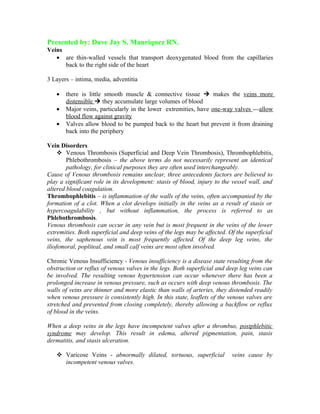
  • 1. Presented by: Dave Jay S. Manriquez RN. Veins • are thin-walled vessels that transport deoxygenated blood from the capillaries back to the right side of the heart 3 Layers – intima, media, adventitia • there is little smooth muscle & connective tissue Ú makes the veins more distensible Ú they accumulate large volumes of blood • Major veins, particularly in the lower extremities, have one-way valves ---allow blood flow against gravity • Valves allow blood to be pumped back to the heart but prevent it from draining back into the periphery Vein Disorders  Venous Thrombosis (Superficial and Deep Vein Thrombosis), Thrombophlebitis, Phlebothrombosis – the above terms do not necessarily represent an identical pathology, for clinical purposes they are often used interchangeably. Cause of Venous thrombosis remains unclear, three antecedents factors are believed to play a significant role in its development: stasis of blood, injury to the vessel wall, and altered blood coagulation. Thrombophlebitis – is inflammation of the walls of the veins, often accompanied by the formation of a clot. When a clot develops initially in the veins as a result of stasis or hypercoagulability , but without inflammation, the process is referred to as Phlebothrombosis. Venous thrombosis can occur in any vein but is most frequent in the veins of the lower extremities. Both superficial and deep veins of the legs may be affected. Of the superficial veins, the saphenous vein is most frequently affected. Of the deep leg veins, the iliofemoral, popliteal, and small calf veins are most often involved. Chronic Venous Insufficiency - Venous insufficiency is a disease state resulting from the obstruction or reflux of venous valves in the legs. Both superficial and deep leg veins can be involved. The resulting venous hypertension can occur whenever there has been a prolonged increase in venous pressure, such as occurs with deep venous thrombosis. The walls of veins are thinner and more elastic than walls of arteries, they distended readily when venous pressure is consistently high. In this state, leaflets of the venous valves are stretched and prevented from closing completely, thereby allowing a backflow or reflux of blood in the veins. When a deep veins in the legs have incompetent valves after a thrombus, postphlebitic syndrome may develop. This result in edema, altered pigmentation, pain, stasis dermatitis, and stasis ulceration.  Varicose Veins - abnormally dilated, tortuous, superficial veins cause by incompetent venous valves.
  • 2. Venous Disorders  alteration in the transport/flow of blood from the capillary back to the heart  changes in smooth muscle and connective tissue make the veins less distensible with limited recoil capacity  valves may malfunction, causing backflow of blood  Virchow’s triad: blood stasis, vessel wall injury, and altered blood coagulation Thrombophlebitis  inflammation of the veins caused by thrombus or blood clot Factors assoc. with the devt. of Thrombophlebitis  venous stasis – occurs when blood flow is retarded, such as heart failure and shock; when veins are dilated, such as after drug therapy; and when skeletal muscle contraction is reduced, as with immobility, extremity paralysis, or anesthesia.  damage to the vessel wall – disruption of the intimal lining of blood vessel creates a site for clot formation, such as after a fracture or dislocation, diseases of the veins, and chemical irritation of the vein from intravenous drugs or solutions.  hypercoagulability of the blood – oral contraceptive use  common to hospitalized pts. , undergone major surgery (pelvic or hip surgery), MI Pathophysiology  develops in both the deep and superficial veins of the lower extremity  deep veins – femoral, popliteal, small calf veins  superficial veins – saphenous vein  Thrombus – form in the veins from accumulation of platelets, fibrin, WBC and RBC Risk Factors for Thrombophlebitis  Bed Rest  General Surgery  Leg Trauma  Previous Venous Insufficiency  Obesity  Oral Contraceptives  Malignancy Deep Vein Thrombosis (DVT)  tends to occur at bifurcations of the deep veins, which are sites of turbulent blood flow  a major risk during the acute phase of thrombophlebitis is dislodgment of the thrombus Ú embolus  pulmonary embolus – is a serious complication arising from DVT of the lower extremities Clinical Manifestations:  pain and edema of extremity – obstruction of venous flow
  • 3.  Û circumference of the thigh or calf  (+) Homan’s sign – dorsiflexion of the foot produces calf pain  Do not check for the Homan’s sign if DVT is already known to be present Ú Û risk of embolus formation  * if superficial veins are affected - signs of inflammation may be noted – redness, warmth, tenderness along the course of the vein, the veins feel hard and thready & sensitive to pressure The risk of dislodgement and embolization of superficial venous thrombi is very low because the majority of them undergo spontaneous lysis, thus this condition can be treated at home with rest, extremity elevation, analgesics, and possibly anti- inflammatory agents. Diagnostic Evaluation Noninvasive Techniques: - rely on the thrombus to create abnormalities of venous flow.  Doppler Ultrasonography – use of a Doppler probe placed over veins that are obstructed. Nonexpensive, portable, simple, rapid, and noninvasive.  Duplex Venous Imaging – able to obtain anatomic information, as well as to assess physiologic parameters.  Impedance Plethysmography – is used to measure changes in venous vlume, a blood pressure cuff is applied to patient thigh inflated enough to impede venous flow (50-60 mmHg), calf electrodes are used to measure electrical resistance that results from venous volume changes. Invasive Techniques: - rely on the injection of contrast media into the venous system, which then bind with structural elements of the thrombus. I-labeled Fibrinogen Scanning - a sensitive method for early detection of venous thrombosis. The test relies on the fact that radioactive fibrinogen, when injected intravenously, will concentrate in the forming clot. The level o radioactivity can then be serially measured by an external counter, and the progression of the clot can be monitored. Contrast Phlebography - involves the injection of radiographic contrast media into the venous system through a dorsal foot vein. Medical Management Superficial thrombophlebitis  bed rest with legs elevated  apply moist heat  NSAID’s ( Non – steroidal anti-inflammatory drugs) - aspirin Deep vein thrombosis  requires hospitalization  bed rest w/ legs elevated to 15-20 degrees above heart level ( knees slightly flexed, trunk horizontal, head may be raised) to promote venous return and help prevent further emboli and prevent edema  application of warm moist heat to reduce pain, promotes venous return  elastic stocking or bandage  anticoagulants, initially with IV heparin then coumadin  fibrinolytic to resolve the thrombus
  • 4.  vasodilator if needed to control vessel spasm and improve circulation Nursing Intervention Preventive care  prevent long periods of standing or sitting that impair venous return  elevate legs when sitting, dorsiflex feet at regular intervals to prevent venous pooling  if edema occurs, elevate above heart level  regular exercise program to promote circulation  avoid crossing legs at the knees  avoid wearing constrictive clothing such as tight bands around socks or garters  use elastic stocking on affected leg  do leg exercises during periods of enforced immobility such as after surgery Nursing Assessment - is invaluable in detecting early signs of venous disorders of the lower extremities. Patients with history of varicose veins, hypercoagulation, neoplastic disease, cardiovascular disease, or recent major surgery or injury, and the obese, the elderly, and women taking oral contraceptives are in the high risk group.  characteristic of the pain  onset & duration of symptoms  history of thrombophlebitis or venous disorders  color & temp. of extremity  edema of calf of thigh - use a tape measure, measure both legs for comparison  Identify areas of tenderness and any thrombosis Surgery  if the thrombus is recurrent and extensive or if the pt. is at high risk for pulmonary embolism - . Surgery for deep vein thrombosis is necessary when: 1. anticoagulant or thrombolytic therapy is contraindicated. 2. the danger of pulmonary embolism is extreme and 3. the venous drainage is so severely compromised that permanent extremity damage will probably result.  Thrombectomy – incising the common femoral vein in the groin and extracting the clots  Vena caval interruption – transvenous placement of a grid or umbrella filter in the vena cava to block the passage of emboli Nursing Management Acute care  explain purpose of bed rest and leg elevation  use elastic stockings  monitor pt. on anticoagulant & fibrinolytic therapy for signs of bleeding  monitor for signs of pulmonary embolism – sudden onset of chest pain, dyspnea, rapid breathing, tachycardia Nsg. intervention often surgery of vena caval interruption  assess insertion site – bleeding, hematoma, apply pressure over site and inform physician
  • 5.  keep pt. on bed rest for 1st 24 hrs. then encourage ROM exercises to promote venous return  assist pt. in ambulation when permitted, elevate legs when sitting  keep elastic bandage  avoid rubbing or massaging the affected extremity  give analgesic and anti-inflammatory agents to promote comfort Anticoagulant Therapy for Thromboembolism: - is the administration of a medication to delay the clotting time of blood, to prevent the formation of a thrombus in postoperative patients, and to forestall the extension of a thrombus once it has formed. Anticoagulants cannot dissolve a thrombus that has already formed. Administration: - Heparin is administered using a continuous pump infusion. To promptly reverse the effects of heparin , the physician may prescribe intravenous injections of protamine sulfate. For Coumadin it is Vitamin K. - Intermittent intravenous injection is another means of administering heparin, in this instance a dilute aqueous solution given every 4 hours. - Oral anticougulants, such as Coumadin, are monitored by the prothrombin time. Because Coumadin has a lag period of 3 to 5 days, it is usually administered in conjunction with heparin until desired anticoagulation has been achieved. Precaution and Nursing Assessment: - The principal complication of anticoagulant therapy is the occurrence of spontaneous bleeding anywhere in the body. A further possible complication of heparin therapy is that of Heparin-induced thrombocytopenia which generally occurs 7 to 10 days after the treatment has been started. Oral anticoagulants interact with many other medications. It is advisory to study drug interactions for patients taking specific oral coagulants. Patient Education About Oral Anticogulants: - The patient should be informed about the medication, its purpose, and the need to take the correct amount at the specific times prescribed, and should be aware that blood tests are scheduled periodically to determine whether a change in medication dosage is required. Chronic Venous Insufficiency  Results from obstruction of venous valves in legs or reflux of blood back through valves  Venous ulceration is serious complication – this is a result of inadequate exchange of oxygen and other nutrients in the tissue, when the cellular metabolism cannot maintain energy balance, cell death (necrosis) results.  Pharmacological therapy is antibiotics for infections  Debridement to promote healing  Topical Therapy may be used with cleansing and debridement
  • 6. Management and Patient Education – management of the patient with venous insufficiency is directed at reducing venous stasis and preventing ulcerations. Measures that increases venous blood flow are antigravity activities and compression of superficial veins with elastic stockings. Elevation of the legs decreases edema, promote venous return. Elevations should be performed frequently throughout the day (at least 30 minutes every 2 hours). At night, the patient should sleep with the foot of the bed elevated for 6 inches. Prolonged sitting or standing still detrimental, but walking should be encouraged. When sitting, patient should avoid placing pressure on the popliteal spaces, such as occurs in leg crossing, or sitting with the legs dangling over the side of the bed. Wearing of constricted garments are also contraindicated. Elastic stockings of the legs reduces pooling of venous blood and enhances venous return to the heart. Prevent twisting of stockings causing tourniquet effect cause this will worsens venous pooling. Extremities with venous insufficiency are conscientiously protected from trauma. The skin is kept clean, dry, and soft. Signs of ulceration are immediately reported to the nurse or physician for treatment and follow-up. Varicose Veins  are abnormally dilated veins with incompetent valves, occurring most often in the lower extremities, the saphenous veins, or the lower trunk however, it can occur elsewhere in the body. Affect1 of 5 persons in the world. This condition is most common in women and in persons in occupations requiring prolonged standing such as salespeople, barbers, beauticians, elevator operators, nurses and dentist. A hereditary weakness of the vein wall may contribute to the development of varicosities, and it is not uncommon to see this condition occur in several members of the same family. • usually affected are woman 30-50 years old. Causes: – congenital absence of a valve – incompetent valves due to external pressure on the veins from pregnancy, ascites or abdominal tumors – sustained Û in venous pressure due to CHF, cirrhosis Prevention – wear elastic stockings during activities that require long standing or when pregnant – moderate exercise, elevation of legs Pathophysiology  the great and small saphenous veins are most often involved weakening of the vein wall does not withstand normal pressureäveins dilate , pooling of bloodävalves become stretched and incompetentämore accumulation of blood in the veins Clinical Manifestations
  • 7.  Primary varicosities – gradual onset and affect superficial veins, appearance of dark tortuous veins. Affected persons may have no symptoms, but cosmetically the appearance of the dilated vein is unappealing. S/sx – dull aches, muscle cramps, pressure, heaviness or fatigue arising from reduced blood flow to the tissues  Secondary Varicosities – affect the deep veins  occur due to chronic venous insufficiency or venous thrombosis S/sx – edema, pain, changes in skin color, ulcerations may occur from venous stasis Diagnostic Evaluation A. Bordie-Trendelenburg test  assess competency of venous valves through measurement of venous filling time  the pt. lies down with the affected leg raised to allow for venous emptying  a tourniquet is then applied above the knee and the pt. stands. The direction and filling time are recorded both before & after the tourniquet is removed  * Incompetent valves are evident when the veins fill rapidly from backward blood flow B. Perthes’ test - is a diagnostic procedure that easily indicates whether the deeper venous system and communicating veins are competent. A tourniquet is applied just below the knee and the patient is asked to walk. If the varicose veins disappear, the deep system and communicating vessels are competent. If the vessels do not empty and become distended on walking, incompetency or obstruction is inferred. C. Additional tests for the presence of Varicose Veins are the Doppler flow meter- detect retrograde flow of blood in superficial veins with incompetent valves after compression of the leg proximally., Phlebography – the injection of radiographic contrast media into the leg veins so that vein anatomy can be visualized during various leg movements. , and Plethysmography – allows measurement of changes in venous blood volume.. Surgical Intervention  indicated or done for prevention or relief of edema, for recurrent leg ulcers or pain or for cosmetic purposes Vein ligation and stripping  the great sapheneous vein is ligated (tied) close to the femoral junction  the veins are stripped out through small incisions at the groin, above & below the knee and at the ankles.  sterile dressing are placed over the incisions and an elastic bandage extending from the foot to the groin is firmly applied DIAGRAM: Ligation and stripping of the great and the small saphenous veins. (A) The tributaries of the saphenous vein have been ligated, and the saphenous vein has been ligated at the saphenofemoral junction . (B) Vein stripper has been inserted from the ankle superiorly to the groin. The vein is stripped from above to downward. A number of alternate incisions may be needed to remove separate varicosities masses. (C) The small
  • 8. saphenous vein is stripped from its junction with the popliteal vein to a point posterior to the lateral malleolus. Sclerotherapy – an irritating chemical (Sotradecol) is injected into the vein, which irritates venous endothelium and produces localized phlebitis and fibrosis, thereby obliterating the lumen. This treatment may be performed alone for small varicosities or may follow vein ligation and stripping. Sclerosing is a palliative not a curative treatment. Nursing care after vein ligation & stripping  Monitor for signs of bleeding, esp. on 1st post-op day  if there is bleeding, elevate the leg, apply pressure over the wound and notify the surgeon  Keep pt. flat on bed for first 4 hrs. after surgery, elevate leg to promote venous return when lying or sitting  Medicate 30 mins. before ambulation and assist patient  Keep elastic bandage snug and intact, do not remove bandage