SlideShare a Scribd company logo
1 of 14
DARK FIELD MICROSCOPY
PRESENATION BY: GIDWANI MANISH N.
1522964
CHARLIE 1
SOURCE: http://www.photomacrography.net/forum/userpix/1469_DSC_4395_2.jpg
CONTENTS.
• WHAT IS DARK FIELD MICROSCOPY?
• HOW STUFF WORKS…?
• TRANSFORMATION.
• TOO EXPENSIVE? HERE’S WHAT YOU CAN DO..
• APPLICATIONS, ADVANTAGES &
DISADVANTAGES.
• EVIDENCE FOR USE IN RESEARCH IMAGING.
• REFERENCES.
CHARLIE 2
WHAT IS DARK FIELD MICROSCOPY?
Dark Field Microscopy is a technique used to
observe unstained samples causing them to
appear brightly lit against a dark, almost purely
black, background.
CHARLIE 3
PIC: Highly magnified image of sugar crystals using darkfield
microscopy technique.
HOW STUFF WORKS…?
CHARLIE 4
SOURCE: http://www.gonda.ucla.edu/bri_core/mic3.gif
LIGHT DIAGRAMS.
CHARLIE
5
SOURCE: http://secure.tutorsglobe.com/CMSImages/1006_phase%20contrast%20microscope.jpg
TRANSFORMATION.
CHARLIE 6
TOO EXPENSIVE? HERE’S WHAT YOU
CAN DO..
• If you do not have access to “STOP” accessories and cannot
afford a dark field kit, there are alternative ways to adapt
your microscope for dark field illumination.
• The expensive stops are all made of opaque material.
• One option is to use a circular object, such as a coin; adhere
the coin to a larger disk and place below the stage.
• You can also cut out a round piece of thick paper, such as
construction paper, cardboard or poster-board, and attach
to the condenser.
• Whatever you use, the trick is to find the right diameter so
that the makeshift stop will block the light and only allow
the oblique rays to illuminate the specimen.
CHARLIE 7
APPLICATIONS.
• Viewing blood cells (biological dark field microscope,
combined with phase contrast)
• Viewing bacteria (biological dark field microscope, often
combined with phase contrast)
• Viewing different types of algae (biological dark field
microscope)
• Viewing hairline metal fractures (metallurgical dark field
microscope)
• Viewing diamonds and other precious stones (gemological
microscope or stereo dark field microscope)
• Viewing shrimp or other invertebrates (stereo dark field
microscope)
CHARLIE 8
ADVANTAGES & DISADVANTAGES.
ADVANTAGES.
• A dark field microscope is ideal for viewing objects
that are unstained, transparent and absorb little or
no light.
• These specimens often have similar refractive
indices as their surroundings, making them hard to
distinguish with other illumination techniques.
• You can use dark field to study marine organisms
such as algae and plankton, diatoms, insects, fibers,
hairs, yeast and protozoa as well as some minerals
and crystals, thin polymers and some ceramics.
• You can also use dark field in the research of live
bacterium, as well as mounted cells and tissues.
• It is more useful in examining external details, such
as outlines, edges, grain boundaries and surface
defects than internal structure.
• Dark field microscopy is often dismissed for more
modern observation techniques such as phase
contrast and DIC, which provide more accurate,
higher contrasted images and can be used to
observe a greater number of specimens.
• Recently, dark field has regained some of its
popularity when combined with other illumination
techniques, such as fluorescence, which widens its
possible employment in certain fields.
DISADVANTAGES.
• First, dark field images are prone to degradation,
distortion and inaccuracies.
• A specimen that is not thin enough or its density differs
across the slide, may appear to have artifacts throughout
the image.
• The preparation and quality of the slides can grossly
affect the contrast and accuracy of a dark field image.
• You need to take special care that the slide, stage, nose
and light source are free from small particles such as dust,
as these will appear as part of the image.
• Similarly, if you need to use oil or water on the condenser
and/or slide, it is almost impossible to avoid all air
bubbles.
• These liquid bubbles will cause images degradation, flare
and distortion and even decrease the contrast and details
of the specimen.
• Dark field needs an intense amount of light to work. This,
coupled with the fact that it relies exclusively on
scattered light rays, can cause glare and distortion.
• It is not a reliable tool to obtain accurate measurements
of specimens.
• Finally, numerous problems can arise when adapting and
using a dark field microscope. The amount and intensity
of light, the position, size and placement of the condenserCHARLIE 9
CHARLIE 10
EVIDENCE FOR USE IN RESEARCH
IMAGING.
CHARLIE 11
SOME MORE EVIDENCE…
CHARLIE 12
REFERENCES.
• http://www.microscopemaster.com/dark-field-microscope.html
• http://public.wsu.edu/~omoto/papers/darkfield.html
• https://www.microscopeworld.com/t-darkfield_microscopy.aspx
• http://www.gonda.ucla.edu/bri_core/mic3.gif
• http://secure.tutorsglobe.com/CMSImages/1006_phase%20contrast%20
microscope.jpg
• http://www.photomacrography.net/forum/userpix/1469_DSC_4395_2.jpg
• Robert M. Macnab (3/5/1976): “Examination of Bacterial Flagellation by
Dark-Field Microscopy.” Journal Of Clinical Microbiology, Sept. 1976, p.
258-265. vol. 4, No. 3.
• Sergiy Patskovsky, Eric Bergeron, David Rioux, and Michel Meunier
(26/6/2014): “Wide-field hyperspectral 3D imaging of functionalized Gold
nanoparticles targeting cancer cells by reflected light microscopy.” J.
Biophotonics 1–7 (2014)/DOI 10.1002/jbio.201400025.
CHARLIE 13
CHARLIE 14

More Related Content

What's hot

Phase contrast microscope
Phase contrast microscopePhase contrast microscope
Phase contrast microscopeNithyaNandapal
 
Dark Field microscopy
Dark Field microscopyDark Field microscopy
Dark Field microscopyRafiaMalik19
 
Electron microscope
Electron microscopeElectron microscope
Electron microscopeFizza Khan
 
Lecture 3 Unit-I Bright-field & Dark-field Microscopy.pdf
Lecture 3 Unit-I Bright-field & Dark-field Microscopy.pdfLecture 3 Unit-I Bright-field & Dark-field Microscopy.pdf
Lecture 3 Unit-I Bright-field & Dark-field Microscopy.pdfSushmitaChoudhary16
 
Phase contrast microscope
Phase contrast microscopePhase contrast microscope
Phase contrast microscopeSalman Khan
 
Various types of microscopes and microscopy
Various types of microscopes and microscopy   Various types of microscopes and microscopy
Various types of microscopes and microscopy Dr.Dinesh Jain
 
Microscopy
MicroscopyMicroscopy
Microscopykarade
 
Phase contrast microscope
Phase contrast microscopePhase contrast microscope
Phase contrast microscopeSuganyaPaulraj
 
Principles and application of light, phase constrast and fluorescence microscope
Principles and application of light, phase constrast and fluorescence microscopePrinciples and application of light, phase constrast and fluorescence microscope
Principles and application of light, phase constrast and fluorescence microscopeMaitriThakor
 
Phase Contrast Microscopy - Microbiology 1st
Phase Contrast Microscopy - Microbiology 1st Phase Contrast Microscopy - Microbiology 1st
Phase Contrast Microscopy - Microbiology 1st RAHUL PAL
 
Phase contrast microscope
Phase contrast microscopePhase contrast microscope
Phase contrast microscopesamharitash
 
Dark ground microscopy
Dark ground microscopyDark ground microscopy
Dark ground microscopy9925752690
 
Electron microscope, principle and application
Electron microscope, principle and applicationElectron microscope, principle and application
Electron microscope, principle and applicationKAUSHAL SAHU
 
Phase contrast and Fluorescent microscope
Phase contrast and Fluorescent microscopePhase contrast and Fluorescent microscope
Phase contrast and Fluorescent microscopeNithiya Pappuraj
 

What's hot (20)

Phase contrast microscope
Phase contrast microscopePhase contrast microscope
Phase contrast microscope
 
Bright field microscopes
Bright field microscopesBright field microscopes
Bright field microscopes
 
Dark Field microscopy
Dark Field microscopyDark Field microscopy
Dark Field microscopy
 
Microscopy
MicroscopyMicroscopy
Microscopy
 
Electron microscope
Electron microscopeElectron microscope
Electron microscope
 
Lecture 3 Unit-I Bright-field & Dark-field Microscopy.pdf
Lecture 3 Unit-I Bright-field & Dark-field Microscopy.pdfLecture 3 Unit-I Bright-field & Dark-field Microscopy.pdf
Lecture 3 Unit-I Bright-field & Dark-field Microscopy.pdf
 
Phase contrast microscope
Phase contrast microscopePhase contrast microscope
Phase contrast microscope
 
Various types of microscopes and microscopy
Various types of microscopes and microscopy   Various types of microscopes and microscopy
Various types of microscopes and microscopy
 
Microscopy
MicroscopyMicroscopy
Microscopy
 
phase contrast microscope
phase contrast microscopephase contrast microscope
phase contrast microscope
 
Phase contrast microscope
Phase contrast microscopePhase contrast microscope
Phase contrast microscope
 
Confocal microscopy
Confocal microscopyConfocal microscopy
Confocal microscopy
 
Principles and application of light, phase constrast and fluorescence microscope
Principles and application of light, phase constrast and fluorescence microscopePrinciples and application of light, phase constrast and fluorescence microscope
Principles and application of light, phase constrast and fluorescence microscope
 
Phase Contrast Microscopy - Microbiology 1st
Phase Contrast Microscopy - Microbiology 1st Phase Contrast Microscopy - Microbiology 1st
Phase Contrast Microscopy - Microbiology 1st
 
Phase contrast microscope
Phase contrast microscopePhase contrast microscope
Phase contrast microscope
 
Dark ground microscopy
Dark ground microscopyDark ground microscopy
Dark ground microscopy
 
Fluorescence microscope
Fluorescence microscopeFluorescence microscope
Fluorescence microscope
 
Light microscope
Light microscopeLight microscope
Light microscope
 
Electron microscope, principle and application
Electron microscope, principle and applicationElectron microscope, principle and application
Electron microscope, principle and application
 
Phase contrast and Fluorescent microscope
Phase contrast and Fluorescent microscopePhase contrast and Fluorescent microscope
Phase contrast and Fluorescent microscope
 

Viewers also liked

Viewers also liked (16)

Cultural characteristics
Cultural characteristicsCultural characteristics
Cultural characteristics
 
History of microbiology
History of microbiologyHistory of microbiology
History of microbiology
 
Conjugation
Conjugation  Conjugation
Conjugation
 
Nitrogen Fixation
Nitrogen FixationNitrogen Fixation
Nitrogen Fixation
 
Blockbusters #soccd
Blockbusters #soccdBlockbusters #soccd
Blockbusters #soccd
 
Transformations
TransformationsTransformations
Transformations
 
History of microbiology
History of microbiologyHistory of microbiology
History of microbiology
 
biodegradation of hydrocarbons
biodegradation of hydrocarbonsbiodegradation of hydrocarbons
biodegradation of hydrocarbons
 
Genetic Analysis and Mapping in Bacteria and Bacteriophages
Genetic Analysis and Mapping in Bacteria and Bacteriophages Genetic Analysis and Mapping in Bacteria and Bacteriophages
Genetic Analysis and Mapping in Bacteria and Bacteriophages
 
Culture characteristic of bacteria
Culture characteristic of bacteriaCulture characteristic of bacteria
Culture characteristic of bacteria
 
Nitrogen cycle
Nitrogen cycleNitrogen cycle
Nitrogen cycle
 
Phase contrast microscope
Phase contrast microscopePhase contrast microscope
Phase contrast microscope
 
NITROGEN CYCLE AND FIXATION
NITROGEN CYCLE AND FIXATIONNITROGEN CYCLE AND FIXATION
NITROGEN CYCLE AND FIXATION
 
Bacterial transformation
Bacterial transformationBacterial transformation
Bacterial transformation
 
Transformation in bacteria
Transformation in bacteriaTransformation in bacteria
Transformation in bacteria
 
Nitrogen fixation
Nitrogen fixationNitrogen fixation
Nitrogen fixation
 

Similar to Dark field microscopy

principlesstructureandapllicationsofbrightfieldanddarkfieldmicroscopy1-copy-2...
principlesstructureandapllicationsofbrightfieldanddarkfieldmicroscopy1-copy-2...principlesstructureandapllicationsofbrightfieldanddarkfieldmicroscopy1-copy-2...
principlesstructureandapllicationsofbrightfieldanddarkfieldmicroscopy1-copy-2...renuka291102
 
Principles, structure and apllications of bright field and dark field microsc...
Principles, structure and apllications of bright field and dark field microsc...Principles, structure and apllications of bright field and dark field microsc...
Principles, structure and apllications of bright field and dark field microsc...selvaraj227
 
Dark Ground Microscopy.pptx
Dark Ground Microscopy.pptxDark Ground Microscopy.pptx
Dark Ground Microscopy.pptxHimaleePachpande
 
Dark Field Microscope (SHOPNIL AKASH)
Dark Field Microscope  (SHOPNIL AKASH)Dark Field Microscope  (SHOPNIL AKASH)
Dark Field Microscope (SHOPNIL AKASH)ShopnilAkash5
 
4-200115131601 (1).pptx
4-200115131601 (1).pptx4-200115131601 (1).pptx
4-200115131601 (1).pptxemadkhan0110
 
3. Dark-field microscopy.pdf
3.             Dark-field microscopy.pdf3.             Dark-field microscopy.pdf
3. Dark-field microscopy.pdfMicrobiologyMicro
 
Dark field Microscope
Dark field MicroscopeDark field Microscope
Dark field MicroscopeArBabIhSan2
 
Microscopy and Types of Microscopes
Microscopy and  Types of MicroscopesMicroscopy and  Types of Microscopes
Microscopy and Types of MicroscopesNCRIMS, Meerut
 
PRELIMINARY RESULTS ON THE PARABOLIC PRIMARY MIRROR MAKING
PRELIMINARY RESULTS ON THE PARABOLIC PRIMARY MIRROR MAKINGPRELIMINARY RESULTS ON THE PARABOLIC PRIMARY MIRROR MAKING
PRELIMINARY RESULTS ON THE PARABOLIC PRIMARY MIRROR MAKINGDhanushka Amaradasa
 
Microscopy and Identification of Mircobes
Microscopy and Identification of MircobesMicroscopy and Identification of Mircobes
Microscopy and Identification of MircobesMuti Ullah Makhmal
 
chapter 2- Microscopy.pptx Microscopy related with medicine
chapter 2- Microscopy.pptx Microscopy related with medicinechapter 2- Microscopy.pptx Microscopy related with medicine
chapter 2- Microscopy.pptx Microscopy related with medicineHarikantSingh4
 

Similar to Dark field microscopy (20)

Dfm
DfmDfm
Dfm
 
principlesstructureandapllicationsofbrightfieldanddarkfieldmicroscopy1-copy-2...
principlesstructureandapllicationsofbrightfieldanddarkfieldmicroscopy1-copy-2...principlesstructureandapllicationsofbrightfieldanddarkfieldmicroscopy1-copy-2...
principlesstructureandapllicationsofbrightfieldanddarkfieldmicroscopy1-copy-2...
 
Principles, structure and apllications of bright field and dark field microsc...
Principles, structure and apllications of bright field and dark field microsc...Principles, structure and apllications of bright field and dark field microsc...
Principles, structure and apllications of bright field and dark field microsc...
 
Dark Ground Microscopy.pptx
Dark Ground Microscopy.pptxDark Ground Microscopy.pptx
Dark Ground Microscopy.pptx
 
Darkfieldmicroscope1
Darkfieldmicroscope1Darkfieldmicroscope1
Darkfieldmicroscope1
 
Dark field microscopy
Dark field microscopyDark field microscopy
Dark field microscopy
 
Dark Field Microscope (SHOPNIL AKASH)
Dark Field Microscope  (SHOPNIL AKASH)Dark Field Microscope  (SHOPNIL AKASH)
Dark Field Microscope (SHOPNIL AKASH)
 
4-200115131601 (1).pptx
4-200115131601 (1).pptx4-200115131601 (1).pptx
4-200115131601 (1).pptx
 
3. Dark-field microscopy.pdf
3.             Dark-field microscopy.pdf3.             Dark-field microscopy.pdf
3. Dark-field microscopy.pdf
 
Dark field Microscope
Dark field MicroscopeDark field Microscope
Dark field Microscope
 
Microscopy and Types of Microscopes
Microscopy and  Types of MicroscopesMicroscopy and  Types of Microscopes
Microscopy and Types of Microscopes
 
Microscope
MicroscopeMicroscope
Microscope
 
Light microscope
Light microscopeLight microscope
Light microscope
 
Presentation1 4
Presentation1 4Presentation1 4
Presentation1 4
 
PRELIMINARY RESULTS ON THE PARABOLIC PRIMARY MIRROR MAKING
PRELIMINARY RESULTS ON THE PARABOLIC PRIMARY MIRROR MAKINGPRELIMINARY RESULTS ON THE PARABOLIC PRIMARY MIRROR MAKING
PRELIMINARY RESULTS ON THE PARABOLIC PRIMARY MIRROR MAKING
 
Microscopy and Identification of Mircobes
Microscopy and Identification of MircobesMicroscopy and Identification of Mircobes
Microscopy and Identification of Mircobes
 
Microscopy
MicroscopyMicroscopy
Microscopy
 
chapter 2- Microscopy.pptx Microscopy related with medicine
chapter 2- Microscopy.pptx Microscopy related with medicinechapter 2- Microscopy.pptx Microscopy related with medicine
chapter 2- Microscopy.pptx Microscopy related with medicine
 
Types of microscopes
Types of microscopes Types of microscopes
Types of microscopes
 
Microscope.pptx
Microscope.pptxMicroscope.pptx
Microscope.pptx
 

Recently uploaded

OECD bibliometric indicators: Selected highlights, April 2024
OECD bibliometric indicators: Selected highlights, April 2024OECD bibliometric indicators: Selected highlights, April 2024
OECD bibliometric indicators: Selected highlights, April 2024innovationoecd
 
final waves properties grade 7 - third quarter
final waves properties grade 7 - third quarterfinal waves properties grade 7 - third quarter
final waves properties grade 7 - third quarterHanHyoKim
 
Dubai Calls Girl Lisa O525547819 Lexi Call Girls In Dubai
Dubai Calls Girl Lisa O525547819 Lexi Call Girls In DubaiDubai Calls Girl Lisa O525547819 Lexi Call Girls In Dubai
Dubai Calls Girl Lisa O525547819 Lexi Call Girls In Dubaikojalkojal131
 
projectile motion, impulse and moment
projectile  motion, impulse  and  momentprojectile  motion, impulse  and  moment
projectile motion, impulse and momentdonamiaquintan2
 
Pests of Bengal gram_Identification_Dr.UPR.pdf
Pests of Bengal gram_Identification_Dr.UPR.pdfPests of Bengal gram_Identification_Dr.UPR.pdf
Pests of Bengal gram_Identification_Dr.UPR.pdfPirithiRaju
 
GENERAL PHYSICS 2 REFRACTION OF LIGHT SENIOR HIGH SCHOOL GENPHYS2.pptx
GENERAL PHYSICS 2 REFRACTION OF LIGHT SENIOR HIGH SCHOOL GENPHYS2.pptxGENERAL PHYSICS 2 REFRACTION OF LIGHT SENIOR HIGH SCHOOL GENPHYS2.pptx
GENERAL PHYSICS 2 REFRACTION OF LIGHT SENIOR HIGH SCHOOL GENPHYS2.pptxRitchAndruAgustin
 
Introduction of Human Body & Structure of cell.pptx
Introduction of Human Body & Structure of cell.pptxIntroduction of Human Body & Structure of cell.pptx
Introduction of Human Body & Structure of cell.pptxMedical College
 
GLYCOSIDES Classification Of GLYCOSIDES Chemical Tests Glycosides
GLYCOSIDES Classification Of GLYCOSIDES  Chemical Tests GlycosidesGLYCOSIDES Classification Of GLYCOSIDES  Chemical Tests Glycosides
GLYCOSIDES Classification Of GLYCOSIDES Chemical Tests GlycosidesNandakishor Bhaurao Deshmukh
 
GenAI talk for Young at Wageningen University & Research (WUR) March 2024
GenAI talk for Young at Wageningen University & Research (WUR) March 2024GenAI talk for Young at Wageningen University & Research (WUR) March 2024
GenAI talk for Young at Wageningen University & Research (WUR) March 2024Jene van der Heide
 
ECG Graph Monitoring with AD8232 ECG Sensor & Arduino.pptx
ECG Graph Monitoring with AD8232 ECG Sensor & Arduino.pptxECG Graph Monitoring with AD8232 ECG Sensor & Arduino.pptx
ECG Graph Monitoring with AD8232 ECG Sensor & Arduino.pptxmaryFF1
 
Forensic limnology of diatoms by Sanjai.pptx
Forensic limnology of diatoms by Sanjai.pptxForensic limnology of diatoms by Sanjai.pptx
Forensic limnology of diatoms by Sanjai.pptxkumarsanjai28051
 
bonjourmadame.tumblr.com bhaskar's girls
bonjourmadame.tumblr.com bhaskar's girlsbonjourmadame.tumblr.com bhaskar's girls
bonjourmadame.tumblr.com bhaskar's girlshansessene
 
trihybrid cross , test cross chi squares
trihybrid cross , test cross chi squarestrihybrid cross , test cross chi squares
trihybrid cross , test cross chi squaresusmanzain586
 
Oxo-Acids of Halogens and their Salts.pptx
Oxo-Acids of Halogens and their Salts.pptxOxo-Acids of Halogens and their Salts.pptx
Oxo-Acids of Halogens and their Salts.pptxfarhanvvdk
 
Call Girls in Majnu Ka Tilla Delhi 🔝9711014705🔝 Genuine
Call Girls in Majnu Ka Tilla Delhi 🔝9711014705🔝 GenuineCall Girls in Majnu Ka Tilla Delhi 🔝9711014705🔝 Genuine
Call Girls in Majnu Ka Tilla Delhi 🔝9711014705🔝 Genuinethapagita
 
Q4-Mod-1c-Quiz-Projectile-333344444.pptx
Q4-Mod-1c-Quiz-Projectile-333344444.pptxQ4-Mod-1c-Quiz-Projectile-333344444.pptx
Q4-Mod-1c-Quiz-Projectile-333344444.pptxtuking87
 
User Guide: Orion™ Weather Station (Columbia Weather Systems)
User Guide: Orion™ Weather Station (Columbia Weather Systems)User Guide: Orion™ Weather Station (Columbia Weather Systems)
User Guide: Orion™ Weather Station (Columbia Weather Systems)Columbia Weather Systems
 
《Queensland毕业文凭-昆士兰大学毕业证成绩单》
《Queensland毕业文凭-昆士兰大学毕业证成绩单》《Queensland毕业文凭-昆士兰大学毕业证成绩单》
《Queensland毕业文凭-昆士兰大学毕业证成绩单》rnrncn29
 
Gas-ExchangeS-in-Plants-and-Animals.pptx
Gas-ExchangeS-in-Plants-and-Animals.pptxGas-ExchangeS-in-Plants-and-Animals.pptx
Gas-ExchangeS-in-Plants-and-Animals.pptxGiovaniTrinidad
 

Recently uploaded (20)

OECD bibliometric indicators: Selected highlights, April 2024
OECD bibliometric indicators: Selected highlights, April 2024OECD bibliometric indicators: Selected highlights, April 2024
OECD bibliometric indicators: Selected highlights, April 2024
 
final waves properties grade 7 - third quarter
final waves properties grade 7 - third quarterfinal waves properties grade 7 - third quarter
final waves properties grade 7 - third quarter
 
Dubai Calls Girl Lisa O525547819 Lexi Call Girls In Dubai
Dubai Calls Girl Lisa O525547819 Lexi Call Girls In DubaiDubai Calls Girl Lisa O525547819 Lexi Call Girls In Dubai
Dubai Calls Girl Lisa O525547819 Lexi Call Girls In Dubai
 
projectile motion, impulse and moment
projectile  motion, impulse  and  momentprojectile  motion, impulse  and  moment
projectile motion, impulse and moment
 
Pests of Bengal gram_Identification_Dr.UPR.pdf
Pests of Bengal gram_Identification_Dr.UPR.pdfPests of Bengal gram_Identification_Dr.UPR.pdf
Pests of Bengal gram_Identification_Dr.UPR.pdf
 
GENERAL PHYSICS 2 REFRACTION OF LIGHT SENIOR HIGH SCHOOL GENPHYS2.pptx
GENERAL PHYSICS 2 REFRACTION OF LIGHT SENIOR HIGH SCHOOL GENPHYS2.pptxGENERAL PHYSICS 2 REFRACTION OF LIGHT SENIOR HIGH SCHOOL GENPHYS2.pptx
GENERAL PHYSICS 2 REFRACTION OF LIGHT SENIOR HIGH SCHOOL GENPHYS2.pptx
 
Introduction of Human Body & Structure of cell.pptx
Introduction of Human Body & Structure of cell.pptxIntroduction of Human Body & Structure of cell.pptx
Introduction of Human Body & Structure of cell.pptx
 
GLYCOSIDES Classification Of GLYCOSIDES Chemical Tests Glycosides
GLYCOSIDES Classification Of GLYCOSIDES  Chemical Tests GlycosidesGLYCOSIDES Classification Of GLYCOSIDES  Chemical Tests Glycosides
GLYCOSIDES Classification Of GLYCOSIDES Chemical Tests Glycosides
 
GenAI talk for Young at Wageningen University & Research (WUR) March 2024
GenAI talk for Young at Wageningen University & Research (WUR) March 2024GenAI talk for Young at Wageningen University & Research (WUR) March 2024
GenAI talk for Young at Wageningen University & Research (WUR) March 2024
 
ECG Graph Monitoring with AD8232 ECG Sensor & Arduino.pptx
ECG Graph Monitoring with AD8232 ECG Sensor & Arduino.pptxECG Graph Monitoring with AD8232 ECG Sensor & Arduino.pptx
ECG Graph Monitoring with AD8232 ECG Sensor & Arduino.pptx
 
Forensic limnology of diatoms by Sanjai.pptx
Forensic limnology of diatoms by Sanjai.pptxForensic limnology of diatoms by Sanjai.pptx
Forensic limnology of diatoms by Sanjai.pptx
 
bonjourmadame.tumblr.com bhaskar's girls
bonjourmadame.tumblr.com bhaskar's girlsbonjourmadame.tumblr.com bhaskar's girls
bonjourmadame.tumblr.com bhaskar's girls
 
trihybrid cross , test cross chi squares
trihybrid cross , test cross chi squarestrihybrid cross , test cross chi squares
trihybrid cross , test cross chi squares
 
Oxo-Acids of Halogens and their Salts.pptx
Oxo-Acids of Halogens and their Salts.pptxOxo-Acids of Halogens and their Salts.pptx
Oxo-Acids of Halogens and their Salts.pptx
 
Call Girls in Majnu Ka Tilla Delhi 🔝9711014705🔝 Genuine
Call Girls in Majnu Ka Tilla Delhi 🔝9711014705🔝 GenuineCall Girls in Majnu Ka Tilla Delhi 🔝9711014705🔝 Genuine
Call Girls in Majnu Ka Tilla Delhi 🔝9711014705🔝 Genuine
 
Q4-Mod-1c-Quiz-Projectile-333344444.pptx
Q4-Mod-1c-Quiz-Projectile-333344444.pptxQ4-Mod-1c-Quiz-Projectile-333344444.pptx
Q4-Mod-1c-Quiz-Projectile-333344444.pptx
 
User Guide: Orion™ Weather Station (Columbia Weather Systems)
User Guide: Orion™ Weather Station (Columbia Weather Systems)User Guide: Orion™ Weather Station (Columbia Weather Systems)
User Guide: Orion™ Weather Station (Columbia Weather Systems)
 
Interferons.pptx.
Interferons.pptx.Interferons.pptx.
Interferons.pptx.
 
《Queensland毕业文凭-昆士兰大学毕业证成绩单》
《Queensland毕业文凭-昆士兰大学毕业证成绩单》《Queensland毕业文凭-昆士兰大学毕业证成绩单》
《Queensland毕业文凭-昆士兰大学毕业证成绩单》
 
Gas-ExchangeS-in-Plants-and-Animals.pptx
Gas-ExchangeS-in-Plants-and-Animals.pptxGas-ExchangeS-in-Plants-and-Animals.pptx
Gas-ExchangeS-in-Plants-and-Animals.pptx
 

Dark field microscopy

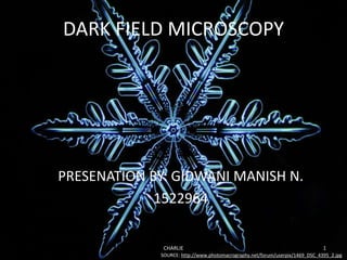
  • 1. DARK FIELD MICROSCOPY PRESENATION BY: GIDWANI MANISH N. 1522964 CHARLIE 1 SOURCE: http://www.photomacrography.net/forum/userpix/1469_DSC_4395_2.jpg
  • 2. CONTENTS. • WHAT IS DARK FIELD MICROSCOPY? • HOW STUFF WORKS…? • TRANSFORMATION. • TOO EXPENSIVE? HERE’S WHAT YOU CAN DO.. • APPLICATIONS, ADVANTAGES & DISADVANTAGES. • EVIDENCE FOR USE IN RESEARCH IMAGING. • REFERENCES. CHARLIE 2
  • 3. WHAT IS DARK FIELD MICROSCOPY? Dark Field Microscopy is a technique used to observe unstained samples causing them to appear brightly lit against a dark, almost purely black, background. CHARLIE 3 PIC: Highly magnified image of sugar crystals using darkfield microscopy technique.
  • 4. HOW STUFF WORKS…? CHARLIE 4 SOURCE: http://www.gonda.ucla.edu/bri_core/mic3.gif
  • 7. TOO EXPENSIVE? HERE’S WHAT YOU CAN DO.. • If you do not have access to “STOP” accessories and cannot afford a dark field kit, there are alternative ways to adapt your microscope for dark field illumination. • The expensive stops are all made of opaque material. • One option is to use a circular object, such as a coin; adhere the coin to a larger disk and place below the stage. • You can also cut out a round piece of thick paper, such as construction paper, cardboard or poster-board, and attach to the condenser. • Whatever you use, the trick is to find the right diameter so that the makeshift stop will block the light and only allow the oblique rays to illuminate the specimen. CHARLIE 7
  • 8. APPLICATIONS. • Viewing blood cells (biological dark field microscope, combined with phase contrast) • Viewing bacteria (biological dark field microscope, often combined with phase contrast) • Viewing different types of algae (biological dark field microscope) • Viewing hairline metal fractures (metallurgical dark field microscope) • Viewing diamonds and other precious stones (gemological microscope or stereo dark field microscope) • Viewing shrimp or other invertebrates (stereo dark field microscope) CHARLIE 8
  • 9. ADVANTAGES & DISADVANTAGES. ADVANTAGES. • A dark field microscope is ideal for viewing objects that are unstained, transparent and absorb little or no light. • These specimens often have similar refractive indices as their surroundings, making them hard to distinguish with other illumination techniques. • You can use dark field to study marine organisms such as algae and plankton, diatoms, insects, fibers, hairs, yeast and protozoa as well as some minerals and crystals, thin polymers and some ceramics. • You can also use dark field in the research of live bacterium, as well as mounted cells and tissues. • It is more useful in examining external details, such as outlines, edges, grain boundaries and surface defects than internal structure. • Dark field microscopy is often dismissed for more modern observation techniques such as phase contrast and DIC, which provide more accurate, higher contrasted images and can be used to observe a greater number of specimens. • Recently, dark field has regained some of its popularity when combined with other illumination techniques, such as fluorescence, which widens its possible employment in certain fields. DISADVANTAGES. • First, dark field images are prone to degradation, distortion and inaccuracies. • A specimen that is not thin enough or its density differs across the slide, may appear to have artifacts throughout the image. • The preparation and quality of the slides can grossly affect the contrast and accuracy of a dark field image. • You need to take special care that the slide, stage, nose and light source are free from small particles such as dust, as these will appear as part of the image. • Similarly, if you need to use oil or water on the condenser and/or slide, it is almost impossible to avoid all air bubbles. • These liquid bubbles will cause images degradation, flare and distortion and even decrease the contrast and details of the specimen. • Dark field needs an intense amount of light to work. This, coupled with the fact that it relies exclusively on scattered light rays, can cause glare and distortion. • It is not a reliable tool to obtain accurate measurements of specimens. • Finally, numerous problems can arise when adapting and using a dark field microscope. The amount and intensity of light, the position, size and placement of the condenserCHARLIE 9
  • 11. EVIDENCE FOR USE IN RESEARCH IMAGING. CHARLIE 11
  • 13. REFERENCES. • http://www.microscopemaster.com/dark-field-microscope.html • http://public.wsu.edu/~omoto/papers/darkfield.html • https://www.microscopeworld.com/t-darkfield_microscopy.aspx • http://www.gonda.ucla.edu/bri_core/mic3.gif • http://secure.tutorsglobe.com/CMSImages/1006_phase%20contrast%20 microscope.jpg • http://www.photomacrography.net/forum/userpix/1469_DSC_4395_2.jpg • Robert M. Macnab (3/5/1976): “Examination of Bacterial Flagellation by Dark-Field Microscopy.” Journal Of Clinical Microbiology, Sept. 1976, p. 258-265. vol. 4, No. 3. • Sergiy Patskovsky, Eric Bergeron, David Rioux, and Michel Meunier (26/6/2014): “Wide-field hyperspectral 3D imaging of functionalized Gold nanoparticles targeting cancer cells by reflected light microscopy.” J. Biophotonics 1–7 (2014)/DOI 10.1002/jbio.201400025. CHARLIE 13

Editor's Notes

  1. When light hits an object, rays are scattered in all azimuths or directions. The design of the dark field microscope is such that it removes the dispersed light, or zeroth order, so that only the scattered beams hit the sample.The introduction of a condenser and/or stop below the stage ensures that these light rays will hit the specimen at different angles, rather than as a direct light source above/below the object. The result is a “cone of light” where rays are diffracted, reflected and/or refracted off the object, ultimately, allowing you to view a specimen in dark field.
  2. Darkfield microscopy relies on a different illumination system. Rather than illuminating the sample with a filled cone of light, the condenser is designed to form a hollow cone of light. The light at the apex of the cone is focused at the plane of the specimen; as this light moves past the specimen plane it spreads again into a hollow cone. The objective lens sits in the dark hollow of this cone; although the light travels around and past the objective lens, no rays enter it The entire field appears dark when there is no sample on the microscope stage; thus the name darkfield microscopy. When a sample is on the stage, the light at the apex of the cone strikes it. The image is made only by those rays scattered by the sample and captured in the objective lens (note the rays scattered by the specimen in Figure 1). The image appears bright against the dark background. This situation can be compared to the glittery appearance of dust particles in a dark room illuminated by strong shafts of light coming in through a side window. The dust particles are very small, but are easily seen when they scatter the light rays. This is the working principle of darkfield microscopy and explains how the image of low contrast material is created: an object will be seen against a dark background if it scatters light which is captured with the proper device such as an objective lens.
  3. 1.Light enters the microscope for illumination of the sample. 2.A specially sized disc, the patch stop (see figure) blocks some light from the light source, leaving an outer ring of illumination. A wide phase annulus can also be reasonably substituted at low magnification. 3.The condenser lens focuses the light towards the sample. 4.The light enters the sample. Most is directly transmitted, while some is scattered from the sample. 5.The scattered light enters the objective lens, while the directly transmitted light simply misses the lens and is not collected due to a direct illumination block (see figure). 6.Only the scattered light goes on to produce the image, while the directly transmitted light is omitted.
  4. If a microscope has built-in elements to easily modify for dark field illumination, the manufacturer usually lists this amongst the observation specifications.You can achieve dark field by using condensers, mirrors and/or a “stop.” Some microscopes come with these accessories or researchers can purchase dark field kits, or even use some common items to adapt a microscope for dark field illumination.In bright field illumination, the object is lit from below the stage, resulting in a larger, contrasted image that can be studied.A dark field microscope blocks this central light with a condenser so that only oblique rays hit the object.An Abbe condenser, for example, contains a concave orb that collects light rays in all azimuths that bounce off a sample to form a cone of illumination.If there is nothing on the stage, the aperture of the condenser is greater than the objective and the view will be completely black.A stop is an opaque object that blocks the central light when placed underneath the stage condenser.This also causes light to scatter in all azimuths, resulting in a cone of light that allows for dark field observation.
  5. The above image, taken by Dr. Arlene Wechezak, won second place. The photographed specimen is the red algae Scagelia, showing reproductive spores and golden diatoms. The technique used to take the shot, dark field microscopy, removes the background by illuminating the sample with light that won’t be collected by the objective lens, creating a very dark background with the sample illuminated in the foreground.
  6. LEFT:(A) S. typhimurium during translational movement. Theperitrichous flagella form apolar bundle (see cartoon, Fig. 1B) which could be mistaken forpolarflagellation. Classifications should therefore be based on the appearance offlagella on stationary cells. Dark-fieldphotomicrograph taken with high-intensity pulsed xenon arc. Bar equals 5 gm. RIGHT: Polar flagellation in pseudomonads. (A) Stationary cell ofP. stutzeri. (B) Same negative [overex- posed duringprinting] to show cell outline. (C) Cartoon combining information from (A) and (B). (D-F) As for (A-C) but with P. aeruginosa (note presence oftwo flagella at pole, although species is predominantly monotrichous). Bar equals 5 ,um.
  7. LEFT: Microscopy images of fixedCD44+-MDA-MB-231 cells incubatedwith CD44-targetedAuNPs taken at differ-ent planes along the optical z-axis (1 μm distance step) in darkfield illumination mode (A1–4) and in reflected light imagingmode (B1–4). For better clarity an embossing filter is applied for imagesA2,A4, B2, B4. RIGHT: