Glucosa (spinreact)
•
2 likes•5,348 views

Este documento describe un método para determinar la concentración de glucosa en muestras de suero o plasma. La glucosa oxidasa cataliza la oxidación de la glucosa a ácido glucónico, produciendo peróxido de hidrógeno. El peróxido de hidrógeno se detecta mediante una reacción cromogénica que produce un color cuya intensidad depende de la concentración de glucosa. El método proporciona valores de glucosa en un rango de 0,04 a 500 mg/dL con buena precisión y exactitud.

Recommended
More Related Content
What's hot
What's hot (20)
Viewers also liked
Viewers also liked (20)
Similar to Glucosa (spinreact)
Similar to Glucosa (spinreact) (20)
Recently uploaded
Recently uploaded (20)
Glucosa (spinreact)
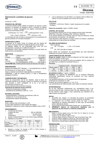
- 1. Glucosa Trinder. GOD-POD BSIS17-E Ed.2007 SPINREACT,S.A.U. Ctra.Santa Coloma, 7 E-17176 SANT ESTEVE DE BAS (GI) SPAIN Tel. +34 972 69 08 00 Fax +34 972 69 00 99. e-mail: spinreact@spinreact.com GLUCOSE -TR Determinación cuantitativa de glucosa IVD Conservar a 2-8ºC PRINCIPIO DEL MÉTODO La glucosa oxidasa (GOD) cataliza la oxidación de glucosa a ácido glucónico. El peróxido de hidrógeno (H2O2), producido se detecta mediante un aceptor cromogénico de oxigeno, fenol-ampirona en presencia de peroxidasa (POD): -D-Glucosa + O2 + H2O GOD Ácido glucónico + H2O2 H2O2 + Fenol + Ampirona POD Quinona + H2O La intensidad del color formado es proporcional a la concentración de glucosa presente en la muestra ensayada 1,2 . SIGNIFICADO CLÍNICO La glucosa es la mayor fuente de energía para las células del organismo; la insulina facilita la entrada de glucosa en las células. La diabetes mellitus es una enfermedad que cursa con una hiperglucémia, causada por un déficit de insulina 1,5,6 . El diagnóstico clínico debe realizarse teniendo en cuenta todos los datos clínicos y de laboratorio. REACTIVOS R 1 Tampón TRIS pH 7,4 Fenol 92 mmol/L 0,3 mmol/L R 2 Enzimas Glucosa oxidasa (GOD) Peroxidasa (POD) 4 - Aminofenazona (4-AF) 15000 U/L 1000 U/L 2,6 mmol/L GLUCOSE CAL Patrón primario acuoso de Glucosa 100 mg/dL PREPARACIÓN Reactivo de trabajo (RT): Disolver ( ) el contenido de un vial de R 2 Enzimas en un frasco de R 1 Tampón. Tapar y mezclar suavemente hasta disolver su contenido. Estabilidad: 1 mes en nevera (2-8ºC) o 7 días a Temperatura ambiente (15-25ºC). CONSERVACIÓN Y ESTABILIDAD Todos los componentes del kit son estables, hasta la fecha de caducidad indicada en la etiqueta, cuando se mantienen los frascos bien cerrados a 2-8ºC, protegidos de la luz y se evita la contaminación durante su uso. No usar reactivos fuera de la fecha indicada. Indicadores de deterioro de los reactivos: - Presencia de partículas y turbidez. - Absorbancia (A) del Blanco a 505 nm 0,10. MATERIAL ADICIONAL - Espectrofotómetro o analizador para lecturas a 505 nm. - Cubetas de 1,0 cm de paso de luz. - Equipamiento habitual de laboratorio. MUESTRAS Suero o plasma, libre de hemólisis 1 y LCR. El suero debe separarse lo antes posible del coágulo. Estabilidad: La glucosa en suero o plasma es estable 3 días a 2-8ºC. PROCEDIMIENTO 1. Condiciones del ensayo: Longitud de onda: . . . . . . . . . . . . . . . 505 nm (490-550) Cubeta:. . . . . . . . . . . . . . . . . . . . . . . .. .1 cm paso de luz Temperatura. . . . . . . . . . . . . . . . . . . . . . . 37ºC / 15-25ºC 2. Ajustar el espectrofotómetro a cero frente a agua destilada. 3. Pipetear en una cubeta: Blanco Patrón Muestra RT (mL) 1,0 1,0 1,0 Patrón (Nota1,2) ( L) -- 10 -- Muestra ( L) -- -- 10 4. Mezclar e incubar 10 minutos a 37ºC ó 15-20 minutos a temperatura ambiente (15-25ºC). 5. Leer la absorbancia (A) del Patrón y la muestra, frente al Blanco de reactivo. El color es estable como mínimo 30 minutos. CÁLCULOS Patrón)A( Muestra)A( x 100 (Conc. Patrón) = mg/dL de glucosa en la muestra Factor de conversión: mg/dL x 0,0555= mmol/L. CONTROL DE CALIDAD Es conveniente analizar junto con las muestras sueros control valorados: SPINTROL H Normal y Patológico (Ref. 1002120 y 1002210). Si los valores hallados se encuentran fuera del rango de tolerancia, revisar el instrumento, los reactivos y el calibrador. Cada laboratorio debe disponer su propio Control de Calidad y establecer correcciones en el caso de que los controles. VALORES DE REFERENCIA 1 Suero o plasma: 60 – 110 mg/dL 3,33 – 6.10 mmol/L LCR: 60 – 80 % del valor en sangre Estos valores son orientativos. Es recomendable que cada laboratorio establezca sus propios valores de referencia. CARACTERISTICAS DEL METODO Rango de medida: Desde el límite de detección de 0,04 mg/dL hasta el límite de linealidad de 500 mg/dL. Si la concentración es superior al límite de linealidad, diluir la muestra 1/2 con ClNa 9 g/L y multiplicar el resultado final por 2. Precisión: Intraserie (n=20) Interserie (n=20) Media (mg/dL) 96,8 241 98,4 248 SD 0,81 1,43 1,55 3,73 CV (%) 0,83 0,59 1,58 1,50 Sensibilidad analítica: 1 mg/dL = 0,0036 A. Exactitud: Los reactivos de SPINREACT (y) no muestran diferencias sistemáticas significativas cuando se comparan con otros reactivos comerciales (x). Los resultados obtenidos con 50 muestras fueron los siguientes: Coeficiente de correlación (r): 0,99. Ecuación de la recta de regresión: y= 1,0x + 0,12. Las características del método pueden variar según el analizador utilizado. INTERFERENCIAS No se han observado interferencias con: hemoglobina hasta 4 g/L, bilirrubina hasta 20 mg/L, creatinina hasta 100 mg/L, galactosa hasta 1 g/L. Se han descrito varias drogas y otras substancias que interfieren en la determinación de la glucosa 3,4 . NOTAS 1. GLUCOSE CAL: Debido a la naturaleza del producto, es aconsejable tratarlo con sumo cuidado ya que se puede contaminar con facilidad. 2. La calibración con el Patrón acuoso puede dar lugar a errores sistemáticos en métodos automáticos. En este caso, se recomienda utilizar calibradores séricos. 3. Usar puntas de pipeta desechables limpias para su dispensación. 4. SPINREACT dispone de instrucciones detalladas para la aplicación de este reactivo en distintos analizadores. BIBLIOGRAFÍA 1. Kaplan A. Glucose. Kaplan A et al. Clin Chem The C.V. Mosby Co. St Louis. Toronto. Princeton 1984; 1032-1036. 2. Trinder P. Ann Clin Biochem 1969; 6: 24-33. 3. Young DS. Effects of drugs on Clinical Lab. Tests, 4th ed AACC Press, 1995. 4. Young DS. Effects of disease on Clinical Lab. Tests, 4th ed AACC 2001. 5. Burtis A et al. Tietz Textbook of Clinical Chemistry, 3rd ed AACC 1999. 6. Tietz N W et al. Clinical Guide to Laboratory Tests, 3rd ed AACC 1995. PRESENTACIÓN Ref:1001190 4 x 125 mL Ref:1001191 4 x 250 mL Ref:1001192 10 x 50 mL Cont.
- 2. Glucose Trinder. GOD-POD BSIS17-E Ed.2007 SPINREACT,S.A.U. Ctra.Santa Coloma, 7 E-17176 SANT ESTEVE DE BAS (GI) SPAIN Tel. +34 972 69 08 00 Fax +34 972 69 00 99. e-mail: spinreact@spinreact.com GLUCOSE -TR Quantitative determination of glucose IVD Store at 2-8ºC PRINCIPLE OF THE METHOD Glucose oxidase (GOD) catalyses the oxidation of glucose to gluconic acid. The formed hydrogen peroxide (H2O2), is detected by a chromogenic oxygen acceptor, phenol-aminophenazone in the presence of peroxidase (POD): -D-Glucose + O2 + H2O GOD Gluconic acid + H2O2 H2O2 + Phenol + Aminophenazone POD Quinone + H2O The intensity of the color formed is proportional to the glucose concentration in the sample 1,2 . CLINICAL SIGNIFICANCE Glucose is a major source of energy for most cells of the body; insulin facilitates glucose entry into the cells. Diabetes is a disease manifested by hyperglycemia; patients with diabetes demonstrate an inability to produce insulin 1,5,6 . Clinical diagnosis should not be made on a single test result; it should integrate clinical and other laboratory data. REAGENTS R 1 Buffer TRIS pH 7.4 Phenol 92 mmol/L 0.3 mmol/L R 2 Enzymes Glucose oxidase (GOD) Peroxidase (POD) 4 – Aminophenazone (4-AP) 15000 U/L 1000 U/L 2.6 mmol/L GLUCOSE CAL Glucose aqueous primary standard 100 mg/dL PREPARATION Working reagent (WR): Dissolve ( ) the contents of one vial R 2 Enzymes in one bottle of R 1 Buffer. Cap and mix gently to dissolve contents. The reagent is stable 1 month after reconstitution in the refrigerator (2-8ºC) or 7 days at room temperature (15-25ºC). STORAGE AND STABILITY All the components of the kit are stable until the expiration date on the label when stored tightly closed at 2-8ºC, protected from light and contaminations prevented during their use. Do not use reagents over the expiration date. Signs of reagent deterioration: - Presence of particles and turbidity. - Blank absorbance (A) at 505 nm 0.10. ADDITIONAL EQUIPMENT - Spectrophotometer or colorimeter measuring at 505 nm. - Matched cuvettes 1.0 cm light path. - General laboratory equipment. SAMPLES Serum or plasma, free of hemolysis1 and CSF. Serum should be removed from the clot as quickly as possible. Stability: Glucose is stable at 2-8ºC for 3 days. PROCEDURE 1. Assay conditions: Wavelength: . . . . . . . . . . . . . .. . 505 nm (490-550) Cuvette: . . . . . . . . . . . . . . . . . . . . .. 1 cm light path Temperature. . . . . . . . . . . . . . . . . . . 37ºC / 15-25ºC 2. Adjust the instrument to zero with distilled water. 3. Pipette into a cuvette: Blank Standard Sample WR (mL) 1.0 1.0 1.0 Standard (Note 1,2) ( L) -- 10 -- Sample ( L) -- -- 10 4. Mix and incubate for 10 min at 37ºC or 15-20 min at room temperature (15-25ºC). 5. Read the absorbance (A) of the samples and standard, against the Blank. The colour is stable for at least 30 minutes. CALCULATIONS dardtanS)A( Sample)A( x 100 (Standard conc.) = mg/dL glucose in the sample Conversion factor: mg/dL x 0.0555= mmol/L. QUALITY CONTROL Control sera are recommended to monitor the performance of assay procedures: SPINTROL H Normal and Pathologic (Ref. 1002120 and 1002210). If control values are found outside the defined range, check the instrument, reagents and calibrator for problems. Each laboratory should establish its own Quality Control scheme and corrective actions if controls do not meet the acceptable tolerances. REFERENCE VALUES 1 Serum or plasma: 60 – 110 mg/dL 3.33 – 6.10 mmol/L CSF: 60 – 80% of the blood value These values are for orientation purpose; each laboratory should establish its own reference range. PERFORMANCE CHARACTERISTICS Measuring range: From detection limit of 0.04 mg/dL to linearity limit of 500 mg/dL. If the results obtained were greater than linearity limit, dilute the sample 1/2 with NaCl 9 g/L and multiply the result by 2. Precision: Intra-assay (n=20) Inter-assay (n=20) Mean (mg/dL) 96.8 241 98.4 248 SD 0.81 1.43 1.55 3.73 CV (%) 0.83 0.59 1.58 1.50 Sensitivity: 1 mg/dL = 0.0036 A. Accuracy: Results obtained using SPINREACT reagents (y) did not show systematic differences when compared with other commercial reagents (x). The results obtained using 50 samples were the following: Correlation coefficient (r): 0.99. Regression equation: y= 1.0x + 0.12. The results of the performance characteristics depend on the analyzer used. INTERFERENCES Haemoglobin up to 4 g/L, bilirubin up to 20 mg/L, creatinine up to 100 mg/L and galactose up to 1g/L do not interfere. A list of drugs and other interfering substances with glucose determination has been reported by Young et. al 3,4 . NOTES 1. GLUCOSE CAL: Proceed carefully with this product because due its nature it can get contamined easily. 2. Calibration with the aqueous standard may cause a systematic error in automatic procedures. In these cases, it is recommended to use a serum Calibrator. 3. Use clean disposable pipette tips for its dispensation. 4. SPINREACT has instruction sheets for several automatic analyzers. Instructions for many of them are available on request. BIBLIOGRAPHY 1. Kaplan L.A. Glucose. Kaplan A et al. Clin Chem The C.V. Mosby Co. St Louis. Toronto. Princeton 1984; 1032-1036. 2. Trinder P. Ann Clin Biochem 1969; 6: 24-33. 3. Young DS. Effects of drugs on Clinical Lab. Tests, 4th ed AACC Press, 1995. 4. Young DS. Effects of disease on Clinical Lab. Tests, 4th ed AACC 2001. 5. Burtis A et al. Tietz Textbook of Clinical Chemistry, 3rd ed AACC 1999. 6. Tietz N W et al. Clinical Guide to Laboratory Tests, 3rd ed AACC 1995. PACKAGING Ref:1001190 4 x 125 mL Ref:1001191 4 x 250 mL Ref:1001192 10 x 50 mL Cont.