call girls in Connaught Place DELHI 🔝 >༒9540349809 🔝 genuine Escort Service ...
When proteins misbehave (part 3)
1. WHEN PROTEINS MISBEHAVE (Part 3)
Why Study Prion Proteins?
Prion proteins(PrPC) are natural proteins that all of us have in our cells. Brain cells have more prion
proteins than the rest of the body. Prion proteins have a number of physiological roles including
synaptic transmission and neuroprotection. PrPC absence results inincreased neuronal excitability and
enhanced excitotoxicityboth in vitro and in vivo.
A misfolded prion protein becomes infectious and is able to influence other prion proteins to misfold
and become infectious as well.
http://www.youtube.com/watch?v=w5aAPEYIL9A
How is the prion protein neuroprotective?
The exact mechanism by which prion proteins helps neuronal survival is unknown, but some studies
indicate that the PrPC inhibits N-methyl-D-aspartic acid (NMDA)–mediated neurotransmission by
interacting with two glutamate receptor subunits. The prion protein is also known to interact with the
postsynaptic density protein PSD-95 (associated with the NMDA receptor) which plays a fundamental
role in synaptic plasticity.
More informations on prion protein neuroprotection:
http://www.landesbioscience.com/journals/prion/article/19639/
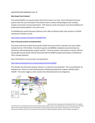
The ultimate role of the prion protein, however is to promote neurite growth. This is accomplished by
binding to the laminin γ1 chain whichactivates metabotropic glutamate receptors mGluR1 and/or
mGluR5. This action triggers a chain reaction that ultimately leads to neuritogenesis.
Schematic model of signaling events mediated by PrPC-Ln γ1 peptide
interaction.PrPC-Ln γ1 peptide interaction activates mGluR1 and/or mGluR5 receptors, promoting activation of PLC, which generates InsP3 that
binds to InsP3 receptors localized to the ER. The activation of InsP3 receptors results in the release of intracellular Ca2+ from the ER and
promotes the activation of PKC and its translocation to the membrane, followed by ERK1/2 activation, which mediates neuritogenesis.
2. The “prion”
Misfolded prion proteins lead to prion diseases which are infectious and deadly. A recent study showed
that a monomeric α-helical form of an amyloidogenic protein is highly cytotoxic. This single molecule or
“monomer” challenges the prevailing concept that neuronal damage is linked to the toxicity of prion
protein aggregates called “oligomers.” By identifying a single molecule as the most toxic species of prion
proteins, a new chapter in our understanding how prion-induced neurodegeneration occurs has begun.
This opens up the possibility that other protein misfolding diseases may be found as well.
http://www.ncbi.nlm.nih.gov/pubmed/22323583
Protein misfolding and neurodegeneration.
For unknown reasons the misfolded proteins are spared by the quality control of the cell Ubiquitin
Proteasome System (UPS). UPS is responsible for catching and recycling incorrectly built or denaturated
proteins, but this mechanism is inefficient in prions and other misfolded proteins. Here is a video of the
physiology of the UBS:
http://www.youtube.com/watch?v=jo8gx61BR-Y
Misfolded Proteins in Diabetes Mellitus Type 2
People with adult onset (type 2) diabetes typically exhibit misfolded, insoluble protein aggregates in
their pancreas. These aggregates damage the membrane of β-cells, hindering the production of insulin.
Research data suggest the involvement of the endoplasmic reticulum (ER) in β-cell survival. β-cells are
the most sensitive to endoplasmic reticulum stress because of the large fluctuations in protein synthesis
they face on a daily basis. When ER homeostasis is disrupted, the ER generates adaptive signaling
3. pathways, called the unfolded protein response (UPR), to maintain homeostasis of this organelle.
However, if homeostasis fails to be restored, the ER initiates death signaling pathways.New observations
suggest that both chronic hyperglycemia and hyperlipidemia, known as important causative factors of
type 2 diabetes (T2D), disrupt ER homeostasis to induce UPR activation and β-cell death
(apoptosis).Chronic ER stress is increasingly being recognized as a factor in many human diseases such
as diabetes, neurodegenerative disorders, and cancer.
ADONIS SFERA, MD