1. Use it or Lose it: Neuroanatomical evolution in response to a changing
environment
BACKGROUND
• Vertebrate brain can evolve in response to environment
• Habitat complexity positively correlated to hippocampus size/volume
in species of birds, bats, and fish (Krebs et al.1989; Safi and Dechmann, 2005; Shumway 2008)
• Little understood concerning:
§ Environmental effects on intraspecific variation in neural-
architecture
§ Sex differences
• The hippocampus is a key structure in vertebrates responsible for
spatial memory and navigation
• In lizards, hippocampal analog reported to be the medial cortex
(Hoogland and Vermeulen-Van der Zee, 1987; Day et al. 2001)
SYSTEM: Lesser Earless Lizard
(Holbrookia maculata)
• Populations undergoing ecological speciation event in two drastically
different habitats in southern New Mexico (Rosenblum and Harmon 2011)
1) Ancestral Chihuahuan Desert dark soil
2) Newer White Sands formation in last 2,000-7,000 years
• Dark Soil quantified as more complex in regards to terrain, competitor
species, and predator species
(Des Roches et al. 2011)
Hypothesis: Habitat
complexity is related
to medial cortex size.
Prediction 1: Lizards
living in a less complex
habitat (White Sands)
will have a reduced medial cortex size
compared to dark soil counterparts.
Prediction 2: Sex differences in medial cortex
size, as males travel more (Jones and Droge 1980)
SIGNIFICANCE
By integrating the fields of neurobiology,
ecology, and evolutionary biology, we have
uncovered differences in neuroanatomy
between habitats and between the sexes in
a wild vertebrate species.
FUTURE DIRECTIONS
• Plasticity at level of individual vs. selective pressure on populations
• Comparative analyses between species with different life strategies
(active foragers vs. sit and wait predators)
• Differences in cell density and morphology contributing to
differences in nucleus area
Sana Chintamen1, Rebecca M. Calisi2,3,4, Lance J. Kriegsfeld4,5, Erica Bree Rosenblum3
1Department of Molecular and Cell Biology, University of California, Berkeley ;2 Department of Biology, Barnard College, Columbia University; 3Department of Environmental Science and Policy
Management, University of California, Berkeley; 4 Department of Psychology, University of California, Berkeley; 5 Helen Wills Neuroscience Institute, University of California, Berkeley
Photographs of two ecomoprhs of H. maculata from both the Chihuahuan Desert
(left) and from White Sands National Monument (right). Photos taken by Simone
Des Roches.
METHODS
MC
• Lizards collected in field, summer
2013
• 20 µm coronal brain sections
• Cresyl violet to stain nuclei
• Area of band of cells from medial
cortex as well as overall brain size
measured with ImageJ software
• 1-tailed t-tests with Welch’s
correction
• Cohen’s d for effect size
RESULTS & DISCUSSION
Hypothesis and Predictions supported
Dark soil lizards, living in more complex
environment, had larger medial cortices than
White Sands lizards
• This is consistent with the idea that in a less complex
environment, there is reduced selective pressure for
maintaining a larger medial cortex
We also found differences in mean medial
cortex area between the sexes
• In the more complex environment (dark soil), males had
larger medial cortices than females
§ consistent with greater roaming behavior in
males and thus potential exposure to more
habitat complexity than females
REFERENCES & ACKNOWLEDGEMENTS
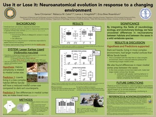
RESULTS
Figure 2: Percent of medial cortex area in relation to total brain area between a) dark soil and White Sands
populations, and b) males and females (pooled across populations).
Figure 3: Percent of medial cortex area in relation to total brain area between males and females of dark soil
and White Sands populations (top), and within dark soil and White Sands populations (bottom).
Figure 4: Histograms depicting relative size of
medial cortex as a function of percentage of
nucleus area to total cortex area of individual
lizards measured. Distribution of relative nucleus
size appears normal.
n=4
n=4
n=5
n=5
REFERENCES:
Day L, Crews D, Wilczynski W (2001): Effects of medial and dorsal cortex lesions on spatial memory in lizards. Behav Brain Res 118:27– 42.
DesRoches, S., L.J. Harmon, E.B. Rosenblum. 2011. Ecological release in White Sands lizards. Ecology and Evolution 1(4): 571-578
Jones, Stephen M., Droge, Dale L. 1980. Home Range Size and Spatial Distributions of Two Sympatric Lizard Species (Sceloporus Undulatus, Holbrookia
Maculata) in the Sand Hills of Nebraska.Herpetologica 36(2):127-132.
Hoogland, P.V., Vermeulen-Van der Zee, E. 1987. Intrinsic and extrinsic connections of the cerebral cortex of lizards.W.K. Schwerdtfeger, W.J.A.J. Smeets
(Eds.), The Forebrain of Reptiles, Karger, Basel, Switzerland, pp. 20–29.
Krebs, J.R., D.F. Sherry, S.D. Healy, V.H. Perry, and A.L. Vaccarino (1989) Hippocampal specialization of food storing birds. Proc. Nat. Acad. Sci. USA, 86: 1388–
1392.
Rosenblum, E.B., L.J. Harmon. 2011. "Same same but different": Replicated ecological speciation at White Sands. Evolution 65(4):946-960.
Safi K, Dechmann D (2005): Adaptations of brain regions to habitat complexity: a comparative analysis in bats (Chiroptera). Proc Biol Sci 272:179–186.
Shumway C (2008): Habitat complexity, brain, and behavior. Brain Behav Evol 72:123–134.
Funding provided by NSF DEB-1054062 to E.B. Rosenblum, & NSF DBI 1003112
& UC President’s Postdoctoral Fellow to R.M. Calisi. Thank you Kayla Hardwick
and Dr. Simone Des Roches for assistance in the field as well as Dr. Benjamin
Smarr, Professor Lance Kriegsfeld, and Professor Erica Bree Rosenblum. A special
thanks to Professor Rebecca Calisi for your constant support and mentorship.
No significant difference between:
• (b) Dark soil females and White Sands females
• (d) White Sands males and White Sands females
Difference between:
• (a) Dark soil males and White Sands males
• (c) Dark soil males and dark soil females
Figure
1:
Illustra3on
(provided
by
Benjamin
Smarr)
depic3ng
plane
sec3oned
(leA)
and
representa3ve
sec3on
stained
with
cresyl
violet
with
stylized
hemisphere
highligh3ng
medial
cortex
(right).