SlideShare a Scribd company logo
1 of 6
Download to read offline
Dengue reporter viruses reveal viral dynamics in
interferon receptor-deficient mice and sensitivity
to interferon effectors in vitro
John W. Schoggins, Marcus Dorner, Michael Feulner, Naoko Imanaka, Mary Y. Murphy, Alexander Ploss,
and Charles M. Rice1
Laboratory of Virology and Infectious Disease, Center for the Study of Hepatitis C, The Rockefeller University, New York, NY 10065
Contributed by Charles M. Rice, July 19, 2012 (sent for review June 16, 2012)
Dengue virus (DENV) is a global disease threat for which there are
no approved antivirals or vaccines. Establishing state-of-the-art
screening systems that rely on fluorescent or luminescent report-
ers may accelerate the development of anti-DENV therapeutics.
However, relatively few reporter DENV platforms exist. Here, we
show that DENV can be genetically engineered to express a green
fluorescent protein or firefly luciferase. Reporter viruses are
infectious in vitro and in vivo and are sensitive to antiviral
compounds, neutralizing antibodies, and interferons. Biolumines-
cence imaging was used to follow the dynamics of DENV infection
in mice and revealed that the virus localized predominantly to
lymphoid and gut-associated tissues. The high-throughput poten-
tial of reporter DENV was demonstrated by screening a library of
more than 350 IFN-stimulated genes for antiviral activity. Several
antiviral effectors were identified, and they targeted DENV at two
distinct life cycle steps. These viruses provide a powerful platform
for applications ranging from validation of vaccine candidates to
antiviral discovery.
in vivo imaging | antiviral screening | flavivirus | interferon-stimulated gene
Dengue virus (DENV) is an arthropod-borne pathogen of the
Flaviviridae family that causes significant morbidity and
mortality worldwide. Each year, over 50 million people are af-
fected by dengue fever, and ∼500,000 are hospitalized with the
more severe dengue hemorrhagic fever (1). The virus is endemic
to tropical environments in Southeast Asia, the Pacific, and the
Americas, and has recently reemerged as far north as southern
Florida (2). Currently, no vaccines or antivirals have been ap-
proved for prevention or treatment of DENV infection.
Four genetically and antigenically distinct DENV serotypes
circulate, and each can be isolated from infected human sera and
propagated in cell culture. The pathogenesis and immune re-
sponse to patient-derived virus can be studied in vitro and in vivo
by quantifying viral genomes or antigens. These detection
methods are also used to study the molecular virology of DENV
with reagents such as virus-like particles (VLPs) and subgenomic
replicons. In some cases, VLPs and subgenomic replicons have
been engineered to take advantage of reporter proteins, such as
luciferase, which can be used in high-throughput screening
platforms for discovery of inhibitors of viral entry or replication
(3). A caveat to these tools is the inability to fully recapitulate the
entire viral life cycle. This can be overcome by launching virus
production from full-length infectious molecular clones, which
have been generated for DENV serotypes 1, 2, and 4 (4–6).
Full-length flavivirus infectious clones are often difficult to
work with, largely due to instability in various bacterial cell lines
and the inability to rescue sufficient quantities of DNA for
downstream applications (7). Additionally, mutagenesis of viral
sequences may result in disruption of the various long-range
interactions required for establishment of a productive replica-
tion complex (8–10). Thus, strategies to generate infectious
viruses expressing heterologous sequences have to contend with
both suboptimal growth conditions in Escherichia coli and the
complex secondary RNA structural requirements of the
viral genome.
Recently, several groups reported the development of in-
fectious serotype 2 DENV (strain TSV01) expressing Renilla
luciferase (11–13). Though this luciferase variant has utility in
a number of applications, such as drug discovery, it is considered
inferior to firefly luciferase with respect to bioluminescence im-
aging (14). We sought to generate infectious DENV based on
serotype 2 strain 16681 that express either GFP or firefly lucif-
erase for cell-based screening assays and bioluminescence im-
aging of DENV infection in mice. Fully infectious viruses
expressing these reporter proteins could be rescued and were
infectious in vitro and in vivo. A luciferase-expressing virus was
used to characterize the dynamics of DENV infection in living
mice, revealing infection primarily in immune organs and gut-
associated tissues. The GFP-expressing recombinant virus was
used as a tool in a cell-based screen to probe a collection of 350+
type I IFN-induced genes for inhibitors of viral replication.
Several anti-DENV effectors were identified, and they selectively
targeted two distinct life cycle stages. These viruses provide
a platform for future screens to identify antiviral molecules and
probe the mechanisms of action of individual IFN effectors
against DENV.
Results
Construction and Characterization of Reporter DENV. To generate
serotype 2-based DENV expressing reporter proteins, we used
the IC30P-A full-length infectious clone of strain 16681 as
a template (5). A series of genetic modifications were made to
enable insertion of sequences for enhanced GFP and firefly lu-
ciferase (Fluc) (Fig. 1A and SI Materials and Methods). To
overcome commonly encountered challenges when modifying
DENV infectious clones, we used MDS42 reduced-genome E.
coli to propagate full-length clones under optimized growth
conditions (Materials and Methods) (15). Rescued plasmid DNA
was used as a template for T7-driven transcription of viral RNAs,
which were electroporated into World Health Organization
(WHO) Vero cells to generate viral stocks. DENV-GFP stocks
were used to infect Vero cells, which were stained by indirect
immunofluorescence for expression of the viral envelope E
protein using the 4G2 monoclonal antibody. Analysis of infected
cells by fluorescence microscopy and flow cytometry revealed
that more than 90% of cells expressing E protein were also
Author contributions: J.W.S., M.D., and C.M.R. designed research; J.W.S., M.D., M.F., N.I.,
and M.Y.M. performed research; M.D. and A.P. contributed new reagents/analytic tools;
J.W.S., M.D., and C.M.R. analyzed data; and J.W.S., M.D., A.P., and C.M.R. wrote
the paper.
The authors declare no conflict of interest.
1
To whom correspondence should be addressed. E-mail: ricec@rockefeller.edu.
This article contains supporting information online at www.pnas.org/lookup/suppl/doi:10.
1073/pnas.1212379109/-/DCSupplemental.
14610–14615 | PNAS | September 4, 2012 | vol. 109 | no. 36 www.pnas.org/cgi/doi/10.1073/pnas.1212379109
positive for GFP (Fig. 1 B and C). We determined that maximum
yields of DENV-GFP were obtained 8 d postelectroporation
using a flow cytometry-based titering assay (Fig. S1 A and B).
Compared with the parental, non-GFP virus, yields of DENV-
GFP were ∼10× lower, as reflected by both infectivity and
titering assays (Fig. 1D). Serially passaging viral supernatants
resulted in diminished GFP fluorescence, demonstrating the
GFP reporter-containing genome is unstable beyond several life
cycles (Fig. S1C), which is consistent with previous reports on
heterologous gene insertion into DENV (11), Sindbis virus (16),
West Nile virus (17), chikungunya virus (18), and influenza virus
(19). Thus, DENV-GFP will be most useful for applications that
monitor single rounds of infection.
A reporter virus expressing firefly luciferase, DENV-Fluc, was
used to demonstrate viral sensitivity to well-characterized anti-
viral compounds mycophenolic acid (MPA), NITD008, and type
I and type III interferons. MPA and NITD008 inhibited DENV-
Fluc with an IC50 of 0.09 μg/mL and 1.5 μM, respectively (Fig.
1E), consistent with previously published studies on nonreporter
DENV (20, 21). DENV-Fluc replication was potently inhibited
by human type I IFN (IFN-α2), and modestly suppressed by
human type III IFNs (IL28B and IL29; Fig. 1F). These data
demonstrate that serotype 2 DENV is amenable to insertion of
a variety of reporter genes, and that heterologous reporters ex-
press at dynamic ranges suitable for testing both chemical and
biological antiviral molecules.
In Vivo Dynamics and Tissue Localization of DENV-Fluc. We next
evaluated the in vivo utility of DENV-Fluc for monitoring viral
dynamics and testing the efficacy of neutralizing antibodies and
antiviral compounds. Using the previously characterized AG129
mouse model of DENV infection (22, 23), which lacks receptors
for IFN-α/IFN-β and IFN-γ, we infected adult mice i.v. with
DENV-Fluc and measured luciferase activity 3 d postinfection
using bioluminescence imaging. Consistent with previous reports
demonstrating that adaptive mutations are needed for DENV to
establish infection even in highly immunocompromised AG129
mice (24), luminescence signal in DENV-Fluc mice did not rise
over the background of an uninfected AG129 mouse (Fig. S2A).
To generate a DENV-Fluc virus suitable for mouse infection, we
took advantage of the recent observation that a single point
mutation (L52F) in the viral nonstrucutural 4B protein (NS4B)
was sufficient to confer virulence to the nonvirulent TSV01 strain
in mice (25). The NS4B L52F mutation was engineered into
DENV-Fluc to generate DENV-Fluc(NS4B:L52F), and both
viruses were characterized for growth and infectivity properties
(Materials and Methods). Maximum titers of both viruses were
obtained 10–11 d after electroporation of viral RNA into Vero
cells (Fig. S3 A and B). When comparing DENV-Fluc(NS4B:
L52F) to DENV-Fluc, we consistently observed a 10- to 100-fold
increase in titers, which correlated to similar increases in lucif-
erase activity. These data indicate that, in cell culture lines, the
NS4B L52F mutation confers a dramatic enhancement in virus
infectivity and production of strain 16681-based DENV-Fluc,
consistent with previous studies on strain TSV01 (25).
To determine if the NS4B point mutation conferred infectivity
in vivo, we injected AG129 mice i.p. with 6 × 10⁵
TCID50
DENV-Fluc(NS4B:L52F) and measured bioluminescence over
6 d. At 24 h, striking Fluc signals were observed in the forelimb
region and on the left side of the main body cavity, consistent
with limb regional lymph nodes and spleen, respectively (Fig. 2 A
and D). At 48 h and 72 h, Fluc signals were variable among in-
dividual mice and typically seen near splenic and gut regions.
When we spatially separated and quantitated Fluc dynamics
across three distinct body regions, we consistently observed ini-
tial infection in the upper body, followed by the appearance of
infection in middle and lower body regions at later time points
(Fig. S2 B–E). Beyond 72 h, luminescence in other anatomical
sites was not observed, and signals eventually reached back-
ground levels across the whole body at day 6 (Fig. 2A). These
kinetic data support previous observations that immune com-
partments are primary sites of DENV infection (26–28), and
highlight the dynamic nature of DENV infection in living mice.
Our data suggest a model in which virus administered i.p. initially
infects lymph nodes near the forelimbs and then disseminates to
the spleen and lower lymph nodes. However, we cannot rule out
the possibility that infection in visceral tissues at later time points
results from a kinetic delay in the onset of replication or is due to
migration of infected cells from upper body lymph nodes.
Given the sharply delineated signals emanating from both
lateral and central lower gut regions, we speculated that in ad-
dition to mesenteric or other lymph nodes, a luciferase signal
might also be emanating from gut associated tissues. A cohort of
six mice was injected with DENV-Fluc(NS4B:L52F), and lym-
phoid and nonlymphoid tissues were harvested for ex vivo lu-
ciferase imaging at 48 h and 72 h postinfection (Fig. 2B). Strong
Fig. 1. Construction and characterization of reporter DENV. (A) Schematic
of the DENV genome expressing heterologous reporter proteins GFP and
Fluc. The first 25 amino acids of DENV capsid were duplicated and placed
upstream of the reporter gene fused to sequences encoding a 2A cleavage
site and ubiquitin. The native cyclization sequence (CS) in full-length capsid
was conservatively mutated to preserved amino acid identity but limit long-
range interactions with the 3′ CS. Asterisk (*) denotes site of L52F mutation
in NS4B. (B) Immunofluorescence microscopy images of WHO Vero cells
infected with DENV-GFP and stained with 4G2 anti-E antibody. (Left) GFP
channel and (Center) immunostaining for E-protein; both panels are gray-
scale and merged in color (Right), with E protein shown as magenta. (C)
FACS plots showing the coexpression of GFP and E protein in cells infected
with DENV-GFP. (D) Comparison of infectivity (left y axis) and titers (right y
axis) of parental DENV or DENV-GFP (left y axis) in Huh7 cells. Data are
presented as mean ± SD, n = 3. (E) Sensitivity of DENV-Fluc to chemical in-
hibition by MPA and NITD008 in Huh7 cells. (F) Sensitivity of DENV-Fluc to
inhibition by type I and III IFNs in Huh7 cells. For E and F, data are presented
as mean ± SD of one of two independent experiments, n = 6.
Schoggins et al. PNAS | September 4, 2012 | vol. 109 | no. 36 | 14611
MICROBIOLOGY
luciferase activity was reproducibly detected in spleen, whereas
signals emanating from heart, brain, lung, and thymus were
similar to those of uninfected animals. In some mice, low lucif-
erase signals were observed in liver and kidney, but this result
was not reproducible across the cohort (Fig. 2B). Surprisingly,
punctae of luminescence were observed in isolated intestines
from all mice at both time points, and signals were more robust
at 72 h (Fig. 2B). These data suggest that gut-associated lym-
phoid tissue may be a target of DENV infection or a destination
for infected migrating cells in this mouse model.
To test whether bioluminescent imaging of DENV-infected
mice could serve as a readout to rapidly identify neutralizing
antibodies or antiviral drugs, we established proof-of-concept
studies with the broadly neutralizing 4G2 monoclonal antibody
and the antiviral compounds MPA and NITD008. Preincubation
of DENV-Fluc(NS4B:L52F) with a neutralizing antibody resulted
in a dose-dependent block to infection in cell culture (Fig. S4A).
Similarly, when the virus was incubated with a 1:10 dilution of 4G2
antibody before infection in mice, we observed Fluc signals that
were similar to uninfected animals at 24 h (Fig. 2C). The adeno-
sine nucleoside inhibitor NITD008 administered at 25 mg/kg body
weight twice daily resulted in a complete block to infection, with
luciferase signals similar to background over a 3-d time course
(Fig. 2D). In this experiment, luciferase signals were markedly
higher than the first time course because we used ∼5× more virus
to demonstrate drug efficacy (compare Fig. 2A with Fig. 2D).
These results complement in vitro data showing potent inhibition
of DENV by NITD008 in cell culture (Fig. 1E). Identical dosing of
MPA did not significantly inhibit infection in mice at early time
points, but provided modest protection at 72 h (Fig. S4C). These
data contrast the strong suppression of DENV replication by
MPA in cell culture (Fig. 1E), suggesting that compounds having
anti-DENV activity in cell-based assays may not necessarily be
effective in mice.
Identification and Characterization of IFN Effectors Targeting DENV-
GFP. In cell culture, DENV is sensitive to type I IFN (29) (Fig.
1F), and type I and II IFN receptor-deficient mice are highly
susceptible to DENV infection (22) (Fig. 2). These observations
suggest that the IFN system contributes significantly to control-
ling DENV replication in vivo. Antiviral IFNs are induced upon
viral infection, signal through the Janus activated kinase/signal
transducer and activator of transcription (JAK-STAT) pathway,
and induce de novo transcription of hundreds of IFN-stimulated
genes (ISGs). The products of these genes facilitate viral clear-
ance and protect uninfected cells from incoming virus. However,
few ISGs with anti-DENV activity have been reported. We re-
cently developed a cell-based expression screen to identify hu-
man antiviral effectors induced by type I IFN (30). The screen
relies on lentiviral delivery of a bicistronic mRNA consisting of
an ISG and TagRFP. ISG-expressing cells are challenged with
a GFP-expressing virus, and replication is quantified by FACS
(Materials and Methods and Fig. S5).
We first determined whether DENV-GFP was suitable for this
screen by testing the IFN effector IFITM3, which was previously
shown to inhibit DENV infection (31). Huh7 cells were trans-
duced with lentiviruses expressing IFITM3 or Fluc as a negative
control. ISG-expressing cells were challenged with DENV-GFP,
and replication was monitored 48 h postinfection by FACS.
IFITM3 robustly inhibited DENV-GFP replication by at least
50% compared with the control Fluc (Fig. 3A). These data
confirm previous findings and validate DENV-GFP as a screen-
ing tool for IFN effector activity.
We next used the DENV-GFP virus to screen a library of more
than 350 ISGs for their ability to inhibit virus replication in hu-
man hepatoma (Huh7) cells. The data are represented by the
level of replication in ISG-expressing cells normalized to Fluc-
expressing control cells (Fig. 3B) and by z score (Fig. S5C). We
found 12 genes that were able to inhibit DENV replication with
a z score of less than −2.0 (Table S1). The hit list includes genes
previously shown to inhibit dengue virus (IFITM2 and IFITM3)
(31), genes that participate in antiviral signaling (RIG-I, IRF1,
IRF7, STAT2, and IL28RA), and genes whose antiviral functions
are either previously unidentified or identified but uncharac-
terized (IFI6, HPSE, NAPA, ADM, and CD9).
To confirm the primary screening hits, we generated in-
dependent lentiviral stocks for nine effectors and tested their
ability to inhibit DENV-GFP in Huh7 cells and immortalized
human STAT1−/−
fibroblasts (32). We also included two genes
(LY6E and MCOLN2) that were previously shown to enhance
the replication of yellow fever virus, a related member of the
Flaviviridae family (30). Most genes were confirmed to inhibit
dengue virus replication to similar levels as the primary screen in
Huh7 (Fig. 3C). In STAT1−/−
fibroblasts, we observed stronger
Fig. 2. Bioluminescent imaging of DENV dynamics, localization, and in-
hibition in vivo. (A and B) Dynamics and localization of AG129 mice injected
with 6 × 10⁵
TCID50 DENV-Fluc(NS4B:L52F). Infected animals were imaged at
indicated time points by injecting 1.5 mg luciferin substrate and monitoring
bioluminescence in the whole animal over a 6-d time course (A) or in isolated
organs at 48 h postinfection (B). (C) Neutralization of DENV infection by
preincubation of 6 × 10⁵
TCID50 DENV-Fluc(NS4B:L52F) with 4G2 anti-DENV
antibody. Signals were quantified with IVIS software. Results are presented
as mean ± SD (n = 10 for mock, n = 8 for DENV, n = 3 for DENV + NAb). (D)
Chemical inhibition of DENV infection with 25 mg/kg body weight NITD008.
Drug was administered upon initial infection with 3 × 10⁶
TCID50 DENV-Fluc
(NS4B:L52F) and every 12 h throughout the course of the experiment. (E)
Quantitation of whole-animal Fluc signals from NITD008 inhibition. Results
are presented as mean ± SD (n = 3 for DENV, n = 4 for DENV + NITD008).
14612 | www.pnas.org/cgi/doi/10.1073/pnas.1212379109 Schoggins et al.
inhibitory or enhancing effects on a per gene basis (Fig. 3C),
consistent with our previous observation that these cells are
more sensitive than Huh7 cells for detecting ISG-mediated
effects (30). Only one gene, NAPA, did not inhibit DENV-GFP
in follow-up assays in Huh7 cells, suggesting a false positive.
However, NAPA expression in STAT1−/−
fibroblasts inhibited
viral replication by ∼30%. Thus, the antiviral properties of this
effector are modest and may depend on the cellular background.
To gain insight into ISG-mediated mechanisms of action with
respect to the virus life cycle, we assessed the kinetics of DENV-
Fluc(NS4B:L52F) replication in STAT1−/−
fibroblasts stably
expressing IFI6, IFITM3, IRF1, STAT2, or an empty cassette.
Stable cell lines were generated by lentiviral transduction followed
by drug selection (SI Materials and Methods). Gene expression of
each ISG was verified by RT–quantitative PCR (Fig. S6A). Using
MPA, which inhibits virus replication but not incoming viral ge-
nome translation (20), we first determined viral kinetics that would
distinguish these two life cycle steps. STAT1−/−
fibroblasts trans-
duced with an empty lentiviral cassette were infected with DENV-
Fluc(NS4B:L52F) and simultaneously treated with MPA or
DMSO control. For the first 24 h of infection, cells treated with
MPA had similar Fluc signals compared with DMSO-treated
control cells (Fig. S4B). After 24 h, MPA robustly inhibited virus
replication, as shown by complete suppression of Fluc signal.
Thus, we chose 12 h as the time point to uncouple the initial round
of translation from later stages of viral replication.
Cell lines stably expressing ISGs were infected with DENV-
Fluc(NS4B:L52F), and Fluc levels were monitored from 12 to 72
h (Fig. 3D). At 12 h, IFITM3 and IRF1 reduced Fluc levels by
more than 50% compared with infected control cells (Fig. 3E).
This level of inhibition indicated a block at or before primary
translation, which is consistent with previous mechanistic studies
on these two genes (30, 31, 33). IFI6- and STAT2-expressing cells
were indistinguishable from control cells at the 12-h time point.
However, these effectors conferred inhibition at 24 h and at later
time points, suggesting a block at the level of genome amplifi-
cation (Fig. 3 F–H). Because IRF1 and STAT2 are both involved
in antiviral signaling, we wanted to determine their contribution
to an antiviral state. We previously showed that IRF1 confers
antiviral activity in a STAT1−/−
background by transcriptionally
inducing a subset of 100–150 ISGs independently of IFN in-
duction (30). To determine if STAT2 confers a similar pheno-
type, we harvested total RNA from cells stably expressing IFI6,
IFITM3, IRF1, and STAT2, and quantified mRNA levels for
ISGs MX1, OAS1, and IFIT1. Only IRF1-expressing cells induced
significant levels of these ISGs over control cells (Fig. S6B).
STAT2 did not confer ISG expression, regardless of whether the
cells were infected with DENV. STAT2 is, therefore, not driving
an antiviral program like IRF1.
Discussion
DENV is a significant global disease threat with few therapeutic
interventions. The successful development of therapeutics for
DENV would likely benefit from viral tools that have robust
readouts for in vitro and in vivo screening. A major hurdle in
dissecting the molecular virology of DENV in the context of
a complete viral life cycle is the relative intractability of full-
length infectious clones. By manipulating the DENV genome
and taking advantage of mouse-adaptive mutations in NS4B, we
have generated unique viruses for monitoring DENV infectivity
in cell culture and in mice. These recombinant viruses have
yielded insight with respect to DENV dynamics in vivo and
susceptibility to IFN effectors.
In this study, we optimized conditions for infectious clone
propagation and engineered fully infectious serotype 2 (strain
16681) DENV expressing heterologous GFP and firefly lucifer-
ase reporters. A key experimental strategy in manipulating
DENV infectious clones was the use of a recently developed E.
coli strain, MDS42. This bacterial line is based on the standard
K-12 E. coli but has 15% of the genome removed, including
nonessential genes and recombinogenic or mobile DNA elements,
which allows propagation of plasmids that may be otherwise un-
stable (15). We found that MDS42 and the RecA-deficient vari-
ant, MDS42Rec, were superior to standard laboratory strains such
as DH5α with respect to faithfully propagating full-length dengue
virus clones. Stbl2 cells could also be used, but total plasmid yields
were lower, suggesting that MDS42-based lines may be superior
for both clone stability and DNA yield.
Fig. 3. Screening reporter DENV against a library of 350+ ISGs. (A) In-
hibition of DENV-GFP by Huh7 cells transduced with lentivirus expressing
IFITM3. Data are represented by FACS plots showing uninfected (gray line)
and infected (black line) cells. (B) Screening 350+ ISGs for antiviral activity
against DENV-GFP. Huh7 cells were transduced with lentiviral stocks for 3 d and
then infected with DENV-GFP. Cells were analyzed for ISG-mediated in-
hibition of DENV-GFP replication by high-throughput FACS. Data are rep-
resented as a dot plot, with replication levels normalized to a Fluc control.
Selected ISGs are indicated in blue. The red line indicates the population
mean. (C) Confirmation assays of select antiviral ISGs in Huh7 (Left) cells or
STAT1−/−
fibroblasts (Right). Replication levels were normalized to the Fluc
control. Data are presented as box and whisker plots: gray boxes extend
from the 25th to the 75th percentile, with a black line at the population
median; whiskers extend to show the highest and lowest values, n = 6. (D)
Time course of ISG-mediated inhibition of DENV-Fluc(NS4B:L52F). ISG-
expressing cells were infected with virus and Fluc levels were monitored over
time. Results are presented as mean ± SD of two independent experiments,
each performed in quadruplicate, n = 8 at each data point. (E–H) Individual
time points of data shown in D. Statistical significance was determined by
one-way ANOVA (***P < 0.001; ns, not significant).
Schoggins et al. PNAS | September 4, 2012 | vol. 109 | no. 36 | 14613
MICROBIOLOGY
DENV reporter viruses were infectious in cell culture and
sensitive to neutralizing antibodies, antiviral compounds, and
antiviral interferons. To study DENV-Fluc infection in living
mice, we incorporated a single amino acid substitution (L52F) in
NS4B. This virus could be grown to high titer and exhibited ro-
bust infectivity in vitro and in vivo. Bioluminescence imaging was
used to probe DENV dynamics in the AG129 mouse model. Our
data suggest that DENV localizes predominantly to lymph nodes
and spleen, but we also observed infection of gut-associated
tissue in all infected animals. Interestingly, the kinetics of lucif-
erase expression indicate that infection may initiate in lymph
nodes and spread to spleen and gut tissues at later time points.
The presence of luciferase punctae in the intestines of animals
infected with DENV-Fluc suggests that DENV can localize to
Peyer’s patches or other gut-associated lymphoid tissues. This
observation is consistent with a recent study on DENV serotype
2 strain S221 infection in AG129 mice (34). In that study, viral
RNA could be detected in the small intestines of infected ani-
mals, and a fraction of isolated laminar propria macrophages
stained positive for DENV prM protein. Further characteriza-
tion of the cell types harboring DENV within these punctae will
be critical for determining whether gut lymphoid tissues play
a role in DENV pathogenesis.
The Fluc-expressing virus was also used to demonstrate sensi-
tivity to a neutralizing antibody and known anti-dengue compounds.
As expected, the adenosine nucleoside inhibitor NITD008 po-
tently suppressed DENV-Fluc replication in cell culture and in
mice. In contrast, mycophenolic acid, a known inhibitor of
DENV replication in cell culture, had little suppressive effect on
the virus in vivo. These data suggest that bioluminescence im-
aging may be useful as an orthogonal screen in drug discovery
pipelines to rapidly identify bioavailable compounds that target
DENV in living animals.
The innate immune system is strongly implicated in the control
of DENV in vivo. To characterize further the nature of innate
immune control on DENV replication, we first confirmed that
our reporter viruses were sensitive to type I and III IFN. To
identify which antiviral effectors may have activity against
DENV, we screened DENV-GFP against a library of more than
350+ ISGs. This screen yielded at least 10 genes that significantly
inhibited viral replication, the majority of which were confirmed
in two distinct cell types. Interestingly, several antiviral effectors
were genes that are known to be involved in antiviral responses
(e.g., IRF1 and IRF7). We previously uncovered each of these
genes while screening other viruses (30), and showed that their
antiviral activity is retained even in a STAT1−/−
cell line. Similar
results were found with DENV, further strengthening the ob-
servation that antiviral effector functions are able to operate
independently of a fully functioning IFN system.
Mechanistically, our studies indicate that IRF1 and IFITM3
target DENV at an early life cycle stage, whereas STAT2 and IFI6
impact later stages of virus replication. Gene expression studies
indicated that STAT2 overexpression is not driving antiviral sig-
naling like IRF1. Previous studies have shown that the DENV
NS5 polymerase potently antagonizes IFN signaling by mediating
STAT2 degradation (35, 36). Our data fit this mechanism and
suggest that STAT2 overexpression may be sufficient to squelch
DENV replication by stoichiometrically overcoming the viral NS5
polymerase. Thus, the STAT2:NS5 interaction may be a unique
target for therapeutic intervention. Moreover, using reporter
DENV to further probe the cellular mechanisms of action of
other antiviral effectors may reveal pathways that can be targeted
for anti-DENV drug discovery.
Materials and Methods
In Vitro Transcription, Virus Production, and Titering. Plasmids bearing full-
length DENV genomes were constructed as described in SI Materials and
Methods. Plasmids were digested with XbaI and the linearized DNA was
used as a template for T7-driven RNA transcription using T7 mMESSAGE
mMACHINE (Ambion) according to the manufacturer’s recommendations
with addition of cap analog. Five micrograms of viral RNAs were electorpo-
rated into WHO Vero cells using a BTX 830 Electroporator (860V, 99-μs pulse
length, five pulses), and virus-containing supernatants were collected 6–13 d
postelectroporation. Virus titration was performed by seeding WHO Vero
cells in poly-L-lysine–coated 96-well plates. Samples were serially diluted
10-fold in complete growth medium and used to infect the seeded cells
(typically 6–8 wells per dilution). Following 2 or 3 d of incubation, the cells were
immunostained for E protein. Wells that expressed at least one E-expressing
cell were counted as positive, and the median tissue culture infective dose
(TCID50) was calculated according to the method of Reed and Muench (37).
For DENV-GFP, infectious unit titers were also determined by limiting dilution
and quantitation of GFP positivity by FACS, as described previously (38).
DENV in Vitro Assays. Typical infection assays were carried out in 24-well plates.
Cell lines were infected in a total volume of 200 μL virus for 2 h at 37 C. After
2 h, 800 μL complete media was added to the cells, and infections proceeded
for a total of 48 h. For indirect immunofluorescence assays, DENV-GFP–
infected cells were stained for E protein with the 4G2 monoclonal antibody
using the Whole Cell Stain kit (Cellomics) according to manufacturer’s
instructions. For DENV-GFP FACS-based assays, media was removed and cells
were detached using 200 μL Accumax Cell Aggregate Dissociation Medium
(eBiosciences). Cells were pelleted by centrifugation, fixed in 1% para-
formaldehyde for at least 20 min, and stored in 1× PBS containing 3% FBS.
GFP fluorescence was monitored by FACS using an LSRII flow cytometer (BD
Biosciences). For DENV-Fluc luciferase-based assays, media was removed and
cells were washed once with 1× PBS before lysis in 1× cell culture lysis buffer
(Promega). Cell lysates were assayed for luciferase activity using the Lucifer-
ase Assay System (Promega). For inhibitor experiments, interferons, drugs, or
DMSO vehicle were added to the virus inoculum and maintained in the media
until the cells were harvested. For antibody inhibition assays, virus inoculum
was incubated on ice with antibody dilutions for 1 h before infection. After a
2-h infection, the antibody-containing inoculum was removed, cells were
washed 1× with PBS, and fresh media was added to the cells.
For ISG inhibition assays, construction and characterization of lentiviral
plasmids and pseudoparticles has been described previously (30). ISG-
expressing lentiviruses were used to transduce Huh7 or STAT1−/−
fibroblasts
by spinoculation. Briefly, 7 × 10⁴
cells were infected with lentiviral pseudo-
particles and transduced by spinning plates at 1,000 × g for 45 min at 37 °C in
media containing 3% FBS, 20 mM Hepes, and 4 μg/mL polybrene. At 48 h
posttransduction, cells were split 1:2 or 1:3. The next day, ISG-expressing cells
were infected with DENV and assayed 48 h later as described above.
DENV in Vivo Assays. Age-matched 129J or AG129 mice were challenged with
DENV-Fluc or DENV-Fluc(NS4B:L52F) at the indicated doses. At regular time
intervals postinfection, mice were anesthetized and injected i.p. with 1.5 mg
luciferin (Caliper Life Sciences). Bioluminescence was measured using an IVIS
Lumina II platform, which was equipped with a supercooled CCD camera
(Caliper Life Sciences; 5-min acquisition time, f/stop 1, binning 4). For antibody
inhibition experiments, virus stocks were preincubated with a 1:10 dilution of
4G2 antibody on ice for 1 h before virus injection. For chemical inhibitor ex-
periments, mice were injected i.p. with virus inoculum containing DMSO vehicle
or 25 mg/kg body weight NITD008 solubilized in DMSO. Mice were dosed with
200 μL 1× PBS containing DMSO vehicle or 25 mg/kg body weight NITD008
every 12 h until the experiment was terminated at 72 h postinfection.
Statistical Analysis. All statistical analyses were performed using the Student t
test, with the exception of ISG confirmation assays, which were analyzed by
one-way ANOVA using Dunnett’s correction.
ACKNOWLEDGMENTS. We thank R. Kinney for the strain 16681 DENV
molecular clone, P.-Y. Shi for the NITD008 compound, E. Jouanguy and J.-L.
Casanova for STAT1−/−
fibroblasts, and S. Wilson and P. Bieniasz for the
SCRPSY-DEST lentiviral plasmid. We also thank S. Pouzol, M. Panis for tech-
nical support; E. Castillo, A. Webson, B. Flatley, and S. Shirley for laboratory
support; R. Labitt for animal care; and The Rockefeller Flow Cytometry Re-
source Center for flow cytometry support. This work was funded in part by
National Institutes of Health Grants AI057158 (Northeast Biodefense Center)
and AI091707 (to C.M.R.). Additional funding was provided by the Green-
berg Medical Research Institute; the Starr Foundation; the Ronald A. Shellow
Memorial Fund (to C.M.R.); National Institute of Diabetes and Digestive and
Kidney Diseases National Research Service Award DK082155 (to J.W.S.);
a postdoctoral fellowship from the German Research Foundation (to M.D.);
and an Astella Young Investigator Award from the Infectious Disease Society
of America (to A.P.).
14614 | www.pnas.org/cgi/doi/10.1073/pnas.1212379109 Schoggins et al.
1. Guzman MG, et al. (2010) Dengue: A continuing global threat. Nat Rev Microbiol 8
(12):Suppl):S7–S16.
2. Centers for Disease Control and Prevention (CDC) (2010) Locally acquired Dengue—
Key West, Florida, 2009-2010. MMWR Morb Mortal Wkly Rep 59:577–581.
3. Noble CG, et al. (2010) Strategies for development of Dengue virus inhibitors. Anti-
viral Res 85:450–462.
4. Puri B, Polo S, Hayes CG, Falgout B (2000) Construction of a full length infectious clone
for dengue-1 virus Western Pacific,74 strain. Virus Genes 20:57–63.
5. Kinney RM, et al. (1997) Construction of infectious cDNA clones for dengue 2 virus:
Strain 16681 and its attenuated vaccine derivative, strain PDK-53. Virology 230:
300–308.
6. Lai CJ, Zhao BT, Hori H, Bray M (1991) Infectious RNA transcribed from stably cloned
full-length cDNA of dengue type 4 virus. Proc Natl Acad Sci USA 88:5139–5143.
7. Rice CM, Grakoui A, Galler R, Chambers TJ (1989) Transcription of infectious yellow
fever RNA from full-length cDNA templates produced by in vitro ligation. New Biol 1:
285–296.
8. Iglesias NG, Gamarnik AV (2011) Dynamic RNA structures in the dengue virus genome.
RNA Biol 8:249–257.
9. Alvarez DE, Lodeiro MF, Ludueña SJ, Pietrasanta LI, Gamarnik AV (2005) Long-range
RNA-RNA interactions circularize the dengue virus genome. J Virol 79:6631–6643.
10. Friebe P, Harris E (2010) Interplay of RNA elements in the dengue virus 5′ and 3′ ends
required for viral RNA replication. J Virol 84:6103–6118.
11. Zou G, Xu HY, Qing M, Wang QY, Shi PY (2011) Development and characterization of
a stable luciferase dengue virus for high-throughput screening. Antiviral Res 91:
11–19.
12. Kaptein SJ, et al. (2010) A derivate of the antibiotic doxorubicin is a selective inhibitor
of dengue and yellow fever virus replication in vitro. Antimicrob Agents Chemother
54:5269–5280.
13. Samsa MM, et al. (2009) Dengue virus capsid protein usurps lipid droplets for viral
particle formation. PLoS Pathog 5:e1000632.
14. Luker KE, Luker GD (2008) Applications of bioluminescence imaging to antiviral re-
search and therapy: Multiple luciferase enzymes and quantitation. Antiviral Res 78:
179–187.
15. Pósfai G, et al. (2006) Emergent properties of reduced-genome Escherichia coli. Sci-
ence 312:1044–1046.
16. Agapov EV, et al. (1998) Noncytopathic Sindbis virus RNA vectors for heterologous
gene expression. Proc Natl Acad Sci USA 95:12989–12994.
17. McGee CE, et al. (2010) Infection, dissemination, and transmission of a West Nile virus
green fluorescent protein infectious clone by Culex pipiens quinquefasciatus mos-
quitoes. Vector Borne Zoonotic Dis 10:267–274.
18. Tsetsarkin K, et al. (2006) Infectious clones of Chikungunya virus (La Réunion isolate)
for vector competence studies. Vector Borne Zoonotic Dis 6:325–337.
19. Manicassamy B, et al. (2010) Analysis of in vivo dynamics of influenza virus infection in
mice using a GFP reporter virus. Proc Natl Acad Sci USA 107:11531–11536.
20. Diamond MS, Zachariah M, Harris E (2002) Mycophenolic acid inhibits dengue virus
infection by preventing replication of viral RNA. Virology 304:211–221.
21. Yin Z, et al. (2009) An adenosine nucleoside inhibitor of dengue virus. Proc Natl Acad
Sci USA 106:20435–20439.
22. Johnson AJ, Roehrig JT (1999) New mouse model for dengue virus vaccine testing. J
Virol 73:783–786.
23. Williams KL, Zompi S, Beatty PR, Harris E (2009) A mouse model for studying dengue
virus pathogenesis and immune response. Ann N Y Acad Sci 1171(Suppl 1):E12–E23.
24. Yauch LE, Shresta S (2008) Mouse models of dengue virus infection and disease.
Antiviral Res 80:87–93.
25. Grant D, et al. (2011) A single amino acid in nonstructural protein NS4B confers vir-
ulence to dengue virus in AG129 mice through enhancement of viral RNA synthesis. J
Virol 85:7775–7787.
26. Pham AM, Langlois RA, TenOever BR (2012) Replication in cells of hematopoietic
origin is necessary for Dengue virus dissemination. PLoS Pathog 8:e1002465.
27. Kyle JL, Beatty PR, Harris E (2007) Dengue virus infects macrophages and dendritic
cells in a mouse model of infection. J Infect Dis 195:1808–1817.
28. Balsitis SJ, et al. (2009) Tropism of dengue virus in mice and humans defined by viral
nonstructural protein 3-specific immunostaining. Am J Trop Med Hyg 80:416–424.
29. Diamond MS, et al. (2000) Modulation of Dengue virus infection in human cells by
alpha, beta, and gamma interferons. J Virol 74:4957–4966.
30. Schoggins JW, et al. (2011) A diverse range of gene products are effectors of the type
I interferon antiviral response. Nature 472:481–485.
31. Brass AL, et al. (2009) The IFITM proteins mediate cellular resistance to influenza A
H1N1 virus, West Nile virus, and dengue virus. Cell 139:1243–1254.
32. Dupuis S, et al. (2003) Impaired response to interferon-alpha/beta and lethal viral
disease in human STAT1 deficiency. Nat Genet 33:388–391.
33. Feeley EM, et al. (2011) IFITM3 inhibits influenza A virus infection by preventing cy-
tosolic entry. PLoS Pathog 7:e1002337.
34. Zellweger RM, Prestwood TR, Shresta S (2010) Enhanced infection of liver sinusoidal
endothelial cells in a mouse model of antibody-induced severe dengue disease. Cell
Host Microbe 7:128–139.
35. Ashour J, Laurent-Rolle M, Shi PY, García-Sastre A (2009) NS5 of dengue virus medi-
ates STAT2 binding and degradation. J Virol 83:5408–5418.
36. Jones M, et al. (2005) Dengue virus inhibits alpha interferon signaling by reducing
STAT2 expression. J Virol 79:5414–5420.
37. Reed LJ, Muench H (1938) A simple method of estimating fifty per cent endpoints. Am
J Epidemiol 72:493–497.
38. Lambeth CR, White LJ, Johnston RE, de Silva AM (2005) Flow cytometry-based assay
for titrating dengue virus. J Clin Microbiol 43:3267–3272.
Schoggins et al. PNAS | September 4, 2012 | vol. 109 | no. 36 | 14615
MICROBIOLOGY

More Related Content

What's hot

New developments in vaccines against African swine fever
New developments in vaccines against African swine feverNew developments in vaccines against African swine fever
New developments in vaccines against African swine feverILRI
 
Towards a vaccine for African swine fever
Towards a vaccine for African swine feverTowards a vaccine for African swine fever
Towards a vaccine for African swine feverILRI
 
SIV Viral Variation; Implications for Vaccines and Transmission - Mars Stone PhD
SIV Viral Variation; Implications for Vaccines and Transmission - Mars Stone PhDSIV Viral Variation; Implications for Vaccines and Transmission - Mars Stone PhD
SIV Viral Variation; Implications for Vaccines and Transmission - Mars Stone PhDMars Stone
 
Current approaches for African swine fever virus vaccine development
Current approaches for African swine fever virus vaccine developmentCurrent approaches for African swine fever virus vaccine development
Current approaches for African swine fever virus vaccine developmentILRI
 
Host response and diagnostic approaches of SARS-COV-2
Host response and diagnostic approaches of SARS-COV-2Host response and diagnostic approaches of SARS-COV-2
Host response and diagnostic approaches of SARS-COV-2Ahmed Al-Abadlah
 
HPV vaccine (UID198612)
HPV vaccine (UID198612)HPV vaccine (UID198612)
HPV vaccine (UID198612)Freya Cardozo
 
Seminario Biología Molecular
Seminario Biología MolecularSeminario Biología Molecular
Seminario Biología MolecularKarollLenT
 
Sigma xi presentation final1
Sigma xi presentation   final1Sigma xi presentation   final1
Sigma xi presentation final1mukta936
 
ProImmune Antigen Characterization Summit Sanja Selak
ProImmune Antigen Characterization Summit Sanja SelakProImmune Antigen Characterization Summit Sanja Selak
ProImmune Antigen Characterization Summit Sanja Selakamandacturner
 
ProImmune Antigen Characterization Summit Gene Olinger
ProImmune Antigen Characterization Summit Gene OlingerProImmune Antigen Characterization Summit Gene Olinger
ProImmune Antigen Characterization Summit Gene Olingeramandacturner
 
Recombinant low-seroprevalent adenoviral vectors Ad26 and Ad35
Recombinant low-seroprevalent adenoviral vectors Ad26 and Ad35Recombinant low-seroprevalent adenoviral vectors Ad26 and Ad35
Recombinant low-seroprevalent adenoviral vectors Ad26 and Ad35Arun kumar
 
Transctiptomic Analysis- Samisti12.ppt
Transctiptomic Analysis- Samisti12.pptTransctiptomic Analysis- Samisti12.ppt
Transctiptomic Analysis- Samisti12.pptFarshad Hamid
 
Sess04 1 jari p.t. valkonen, jan f. kreuze and joseph ndunguru disease mana...
Sess04 1 jari p.t. valkonen, jan f. kreuze and joseph ndunguru   disease mana...Sess04 1 jari p.t. valkonen, jan f. kreuze and joseph ndunguru   disease mana...
Sess04 1 jari p.t. valkonen, jan f. kreuze and joseph ndunguru disease mana...African Potato Association (APA)
 
Genomic surveillance of the Rift Valley fever: From sequencing to Lineage ass...
Genomic surveillance of the Rift Valley fever: From sequencing to Lineage ass...Genomic surveillance of the Rift Valley fever: From sequencing to Lineage ass...
Genomic surveillance of the Rift Valley fever: From sequencing to Lineage ass...ILRI
 
The promise of mRNA vaccines
The promise of mRNA vaccinesThe promise of mRNA vaccines
The promise of mRNA vaccinesZeena Nackerdien
 

What's hot (20)

Scientific Sessions 2015: HIV and genetics
Scientific Sessions 2015: HIV and geneticsScientific Sessions 2015: HIV and genetics
Scientific Sessions 2015: HIV and genetics
 
New developments in vaccines against African swine fever
New developments in vaccines against African swine feverNew developments in vaccines against African swine fever
New developments in vaccines against African swine fever
 
Towards a vaccine for African swine fever
Towards a vaccine for African swine feverTowards a vaccine for African swine fever
Towards a vaccine for African swine fever
 
SIV Viral Variation; Implications for Vaccines and Transmission - Mars Stone PhD
SIV Viral Variation; Implications for Vaccines and Transmission - Mars Stone PhDSIV Viral Variation; Implications for Vaccines and Transmission - Mars Stone PhD
SIV Viral Variation; Implications for Vaccines and Transmission - Mars Stone PhD
 
Current approaches for African swine fever virus vaccine development
Current approaches for African swine fever virus vaccine developmentCurrent approaches for African swine fever virus vaccine development
Current approaches for African swine fever virus vaccine development
 
Public defense
Public defensePublic defense
Public defense
 
2010 phylogeny and serological aspects of the csp p. vivax variants
2010 phylogeny and serological aspects of the csp p. vivax variants2010 phylogeny and serological aspects of the csp p. vivax variants
2010 phylogeny and serological aspects of the csp p. vivax variants
 
Host response and diagnostic approaches of SARS-COV-2
Host response and diagnostic approaches of SARS-COV-2Host response and diagnostic approaches of SARS-COV-2
Host response and diagnostic approaches of SARS-COV-2
 
HPV vaccine (UID198612)
HPV vaccine (UID198612)HPV vaccine (UID198612)
HPV vaccine (UID198612)
 
Seminario Biología Molecular
Seminario Biología MolecularSeminario Biología Molecular
Seminario Biología Molecular
 
Sigma xi presentation final1
Sigma xi presentation   final1Sigma xi presentation   final1
Sigma xi presentation final1
 
ProImmune Antigen Characterization Summit Sanja Selak
ProImmune Antigen Characterization Summit Sanja SelakProImmune Antigen Characterization Summit Sanja Selak
ProImmune Antigen Characterization Summit Sanja Selak
 
ProImmune Antigen Characterization Summit Gene Olinger
ProImmune Antigen Characterization Summit Gene OlingerProImmune Antigen Characterization Summit Gene Olinger
ProImmune Antigen Characterization Summit Gene Olinger
 
Recombinant low-seroprevalent adenoviral vectors Ad26 and Ad35
Recombinant low-seroprevalent adenoviral vectors Ad26 and Ad35Recombinant low-seroprevalent adenoviral vectors Ad26 and Ad35
Recombinant low-seroprevalent adenoviral vectors Ad26 and Ad35
 
Transctiptomic Analysis- Samisti12.ppt
Transctiptomic Analysis- Samisti12.pptTransctiptomic Analysis- Samisti12.ppt
Transctiptomic Analysis- Samisti12.ppt
 
Sess04 1 jari p.t. valkonen, jan f. kreuze and joseph ndunguru disease mana...
Sess04 1 jari p.t. valkonen, jan f. kreuze and joseph ndunguru   disease mana...Sess04 1 jari p.t. valkonen, jan f. kreuze and joseph ndunguru   disease mana...
Sess04 1 jari p.t. valkonen, jan f. kreuze and joseph ndunguru disease mana...
 
Malaria
MalariaMalaria
Malaria
 
Genomic surveillance of the Rift Valley fever: From sequencing to Lineage ass...
Genomic surveillance of the Rift Valley fever: From sequencing to Lineage ass...Genomic surveillance of the Rift Valley fever: From sequencing to Lineage ass...
Genomic surveillance of the Rift Valley fever: From sequencing to Lineage ass...
 
Amer MBT's Defense
Amer MBT's DefenseAmer MBT's Defense
Amer MBT's Defense
 
The promise of mRNA vaccines
The promise of mRNA vaccinesThe promise of mRNA vaccines
The promise of mRNA vaccines
 

Similar to PNAS-2012-Schoggins-14610-5

Detection of dengue viruses using RT-LAMP
Detection of dengue viruses using RT-LAMPDetection of dengue viruses using RT-LAMP
Detection of dengue viruses using RT-LAMPRahul Gupta
 
Dengue Virus: Genomic Insights, Pathogenic Mechanisms, and Therapeutic Approa...
Dengue Virus: Genomic Insights, Pathogenic Mechanisms, and Therapeutic Approa...Dengue Virus: Genomic Insights, Pathogenic Mechanisms, and Therapeutic Approa...
Dengue Virus: Genomic Insights, Pathogenic Mechanisms, and Therapeutic Approa...SindhBiotech
 
Microbiology hiv-yf
Microbiology   hiv-yfMicrobiology   hiv-yf
Microbiology hiv-yfMBBS IMS MSU
 
Human retroviruses
Human retroviruses Human retroviruses
Human retroviruses ankit
 
Mahoney Pipeline Status Siem Reap V2a
Mahoney Pipeline Status Siem Reap V2aMahoney Pipeline Status Siem Reap V2a
Mahoney Pipeline Status Siem Reap V2agambelguy
 
Dengue- WS on vector borne viral infection 2011
Dengue- WS on vector borne viral infection 2011Dengue- WS on vector borne viral infection 2011
Dengue- WS on vector borne viral infection 2011javed.khalil
 
A Mini-Review On Dengue
A Mini-Review On DengueA Mini-Review On Dengue
A Mini-Review On DengueLori Mitchell
 

Similar to PNAS-2012-Schoggins-14610-5 (20)

Advances in Anti-Dengue Virus Research
Advances in Anti-Dengue Virus ResearchAdvances in Anti-Dengue Virus Research
Advances in Anti-Dengue Virus Research
 
AIDS_21(7)_807-811
AIDS_21(7)_807-811AIDS_21(7)_807-811
AIDS_21(7)_807-811
 
HIV/AIDS
HIV/AIDSHIV/AIDS
HIV/AIDS
 
Ebolav2
Ebolav2Ebolav2
Ebolav2
 
Detection of dengue viruses using RT-LAMP
Detection of dengue viruses using RT-LAMPDetection of dengue viruses using RT-LAMP
Detection of dengue viruses using RT-LAMP
 
Dengue Virus: Genomic Insights, Pathogenic Mechanisms, and Therapeutic Approa...
Dengue Virus: Genomic Insights, Pathogenic Mechanisms, and Therapeutic Approa...Dengue Virus: Genomic Insights, Pathogenic Mechanisms, and Therapeutic Approa...
Dengue Virus: Genomic Insights, Pathogenic Mechanisms, and Therapeutic Approa...
 
Tylcv seminar
Tylcv  seminarTylcv  seminar
Tylcv seminar
 
Microbiology hiv-yf
Microbiology   hiv-yfMicrobiology   hiv-yf
Microbiology hiv-yf
 
DF27102016_Rowida
DF27102016_RowidaDF27102016_Rowida
DF27102016_Rowida
 
Animal editing version 3
Animal editing version 3Animal editing version 3
Animal editing version 3
 
Hiv
HivHiv
Hiv
 
Hiv
HivHiv
Hiv
 
Human retroviruses
Human retroviruses Human retroviruses
Human retroviruses
 
Mahoney Pipeline Status Siem Reap V2a
Mahoney Pipeline Status Siem Reap V2aMahoney Pipeline Status Siem Reap V2a
Mahoney Pipeline Status Siem Reap V2a
 
HIV and AIDS /prosthodontic courses
HIV and AIDS /prosthodontic coursesHIV and AIDS /prosthodontic courses
HIV and AIDS /prosthodontic courses
 
Hiv part 1
Hiv part 1Hiv part 1
Hiv part 1
 
Dengue- WS on vector borne viral infection 2011
Dengue- WS on vector borne viral infection 2011Dengue- WS on vector borne viral infection 2011
Dengue- WS on vector borne viral infection 2011
 
Rm2
Rm2Rm2
Rm2
 
Rm2
Rm2Rm2
Rm2
 
A Mini-Review On Dengue
A Mini-Review On DengueA Mini-Review On Dengue
A Mini-Review On Dengue
 

PNAS-2012-Schoggins-14610-5

  • 1. Dengue reporter viruses reveal viral dynamics in interferon receptor-deficient mice and sensitivity to interferon effectors in vitro John W. Schoggins, Marcus Dorner, Michael Feulner, Naoko Imanaka, Mary Y. Murphy, Alexander Ploss, and Charles M. Rice1 Laboratory of Virology and Infectious Disease, Center for the Study of Hepatitis C, The Rockefeller University, New York, NY 10065 Contributed by Charles M. Rice, July 19, 2012 (sent for review June 16, 2012) Dengue virus (DENV) is a global disease threat for which there are no approved antivirals or vaccines. Establishing state-of-the-art screening systems that rely on fluorescent or luminescent report- ers may accelerate the development of anti-DENV therapeutics. However, relatively few reporter DENV platforms exist. Here, we show that DENV can be genetically engineered to express a green fluorescent protein or firefly luciferase. Reporter viruses are infectious in vitro and in vivo and are sensitive to antiviral compounds, neutralizing antibodies, and interferons. Biolumines- cence imaging was used to follow the dynamics of DENV infection in mice and revealed that the virus localized predominantly to lymphoid and gut-associated tissues. The high-throughput poten- tial of reporter DENV was demonstrated by screening a library of more than 350 IFN-stimulated genes for antiviral activity. Several antiviral effectors were identified, and they targeted DENV at two distinct life cycle steps. These viruses provide a powerful platform for applications ranging from validation of vaccine candidates to antiviral discovery. in vivo imaging | antiviral screening | flavivirus | interferon-stimulated gene Dengue virus (DENV) is an arthropod-borne pathogen of the Flaviviridae family that causes significant morbidity and mortality worldwide. Each year, over 50 million people are af- fected by dengue fever, and ∼500,000 are hospitalized with the more severe dengue hemorrhagic fever (1). The virus is endemic to tropical environments in Southeast Asia, the Pacific, and the Americas, and has recently reemerged as far north as southern Florida (2). Currently, no vaccines or antivirals have been ap- proved for prevention or treatment of DENV infection. Four genetically and antigenically distinct DENV serotypes circulate, and each can be isolated from infected human sera and propagated in cell culture. The pathogenesis and immune re- sponse to patient-derived virus can be studied in vitro and in vivo by quantifying viral genomes or antigens. These detection methods are also used to study the molecular virology of DENV with reagents such as virus-like particles (VLPs) and subgenomic replicons. In some cases, VLPs and subgenomic replicons have been engineered to take advantage of reporter proteins, such as luciferase, which can be used in high-throughput screening platforms for discovery of inhibitors of viral entry or replication (3). A caveat to these tools is the inability to fully recapitulate the entire viral life cycle. This can be overcome by launching virus production from full-length infectious molecular clones, which have been generated for DENV serotypes 1, 2, and 4 (4–6). Full-length flavivirus infectious clones are often difficult to work with, largely due to instability in various bacterial cell lines and the inability to rescue sufficient quantities of DNA for downstream applications (7). Additionally, mutagenesis of viral sequences may result in disruption of the various long-range interactions required for establishment of a productive replica- tion complex (8–10). Thus, strategies to generate infectious viruses expressing heterologous sequences have to contend with both suboptimal growth conditions in Escherichia coli and the complex secondary RNA structural requirements of the viral genome. Recently, several groups reported the development of in- fectious serotype 2 DENV (strain TSV01) expressing Renilla luciferase (11–13). Though this luciferase variant has utility in a number of applications, such as drug discovery, it is considered inferior to firefly luciferase with respect to bioluminescence im- aging (14). We sought to generate infectious DENV based on serotype 2 strain 16681 that express either GFP or firefly lucif- erase for cell-based screening assays and bioluminescence im- aging of DENV infection in mice. Fully infectious viruses expressing these reporter proteins could be rescued and were infectious in vitro and in vivo. A luciferase-expressing virus was used to characterize the dynamics of DENV infection in living mice, revealing infection primarily in immune organs and gut- associated tissues. The GFP-expressing recombinant virus was used as a tool in a cell-based screen to probe a collection of 350+ type I IFN-induced genes for inhibitors of viral replication. Several anti-DENV effectors were identified, and they selectively targeted two distinct life cycle stages. These viruses provide a platform for future screens to identify antiviral molecules and probe the mechanisms of action of individual IFN effectors against DENV. Results Construction and Characterization of Reporter DENV. To generate serotype 2-based DENV expressing reporter proteins, we used the IC30P-A full-length infectious clone of strain 16681 as a template (5). A series of genetic modifications were made to enable insertion of sequences for enhanced GFP and firefly lu- ciferase (Fluc) (Fig. 1A and SI Materials and Methods). To overcome commonly encountered challenges when modifying DENV infectious clones, we used MDS42 reduced-genome E. coli to propagate full-length clones under optimized growth conditions (Materials and Methods) (15). Rescued plasmid DNA was used as a template for T7-driven transcription of viral RNAs, which were electroporated into World Health Organization (WHO) Vero cells to generate viral stocks. DENV-GFP stocks were used to infect Vero cells, which were stained by indirect immunofluorescence for expression of the viral envelope E protein using the 4G2 monoclonal antibody. Analysis of infected cells by fluorescence microscopy and flow cytometry revealed that more than 90% of cells expressing E protein were also Author contributions: J.W.S., M.D., and C.M.R. designed research; J.W.S., M.D., M.F., N.I., and M.Y.M. performed research; M.D. and A.P. contributed new reagents/analytic tools; J.W.S., M.D., and C.M.R. analyzed data; and J.W.S., M.D., A.P., and C.M.R. wrote the paper. The authors declare no conflict of interest. 1 To whom correspondence should be addressed. E-mail: ricec@rockefeller.edu. This article contains supporting information online at www.pnas.org/lookup/suppl/doi:10. 1073/pnas.1212379109/-/DCSupplemental. 14610–14615 | PNAS | September 4, 2012 | vol. 109 | no. 36 www.pnas.org/cgi/doi/10.1073/pnas.1212379109
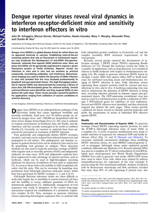
  • 2. positive for GFP (Fig. 1 B and C). We determined that maximum yields of DENV-GFP were obtained 8 d postelectroporation using a flow cytometry-based titering assay (Fig. S1 A and B). Compared with the parental, non-GFP virus, yields of DENV- GFP were ∼10× lower, as reflected by both infectivity and titering assays (Fig. 1D). Serially passaging viral supernatants resulted in diminished GFP fluorescence, demonstrating the GFP reporter-containing genome is unstable beyond several life cycles (Fig. S1C), which is consistent with previous reports on heterologous gene insertion into DENV (11), Sindbis virus (16), West Nile virus (17), chikungunya virus (18), and influenza virus (19). Thus, DENV-GFP will be most useful for applications that monitor single rounds of infection. A reporter virus expressing firefly luciferase, DENV-Fluc, was used to demonstrate viral sensitivity to well-characterized anti- viral compounds mycophenolic acid (MPA), NITD008, and type I and type III interferons. MPA and NITD008 inhibited DENV- Fluc with an IC50 of 0.09 μg/mL and 1.5 μM, respectively (Fig. 1E), consistent with previously published studies on nonreporter DENV (20, 21). DENV-Fluc replication was potently inhibited by human type I IFN (IFN-α2), and modestly suppressed by human type III IFNs (IL28B and IL29; Fig. 1F). These data demonstrate that serotype 2 DENV is amenable to insertion of a variety of reporter genes, and that heterologous reporters ex- press at dynamic ranges suitable for testing both chemical and biological antiviral molecules. In Vivo Dynamics and Tissue Localization of DENV-Fluc. We next evaluated the in vivo utility of DENV-Fluc for monitoring viral dynamics and testing the efficacy of neutralizing antibodies and antiviral compounds. Using the previously characterized AG129 mouse model of DENV infection (22, 23), which lacks receptors for IFN-α/IFN-β and IFN-γ, we infected adult mice i.v. with DENV-Fluc and measured luciferase activity 3 d postinfection using bioluminescence imaging. Consistent with previous reports demonstrating that adaptive mutations are needed for DENV to establish infection even in highly immunocompromised AG129 mice (24), luminescence signal in DENV-Fluc mice did not rise over the background of an uninfected AG129 mouse (Fig. S2A). To generate a DENV-Fluc virus suitable for mouse infection, we took advantage of the recent observation that a single point mutation (L52F) in the viral nonstrucutural 4B protein (NS4B) was sufficient to confer virulence to the nonvirulent TSV01 strain in mice (25). The NS4B L52F mutation was engineered into DENV-Fluc to generate DENV-Fluc(NS4B:L52F), and both viruses were characterized for growth and infectivity properties (Materials and Methods). Maximum titers of both viruses were obtained 10–11 d after electroporation of viral RNA into Vero cells (Fig. S3 A and B). When comparing DENV-Fluc(NS4B: L52F) to DENV-Fluc, we consistently observed a 10- to 100-fold increase in titers, which correlated to similar increases in lucif- erase activity. These data indicate that, in cell culture lines, the NS4B L52F mutation confers a dramatic enhancement in virus infectivity and production of strain 16681-based DENV-Fluc, consistent with previous studies on strain TSV01 (25). To determine if the NS4B point mutation conferred infectivity in vivo, we injected AG129 mice i.p. with 6 × 10⁵ TCID50 DENV-Fluc(NS4B:L52F) and measured bioluminescence over 6 d. At 24 h, striking Fluc signals were observed in the forelimb region and on the left side of the main body cavity, consistent with limb regional lymph nodes and spleen, respectively (Fig. 2 A and D). At 48 h and 72 h, Fluc signals were variable among in- dividual mice and typically seen near splenic and gut regions. When we spatially separated and quantitated Fluc dynamics across three distinct body regions, we consistently observed ini- tial infection in the upper body, followed by the appearance of infection in middle and lower body regions at later time points (Fig. S2 B–E). Beyond 72 h, luminescence in other anatomical sites was not observed, and signals eventually reached back- ground levels across the whole body at day 6 (Fig. 2A). These kinetic data support previous observations that immune com- partments are primary sites of DENV infection (26–28), and highlight the dynamic nature of DENV infection in living mice. Our data suggest a model in which virus administered i.p. initially infects lymph nodes near the forelimbs and then disseminates to the spleen and lower lymph nodes. However, we cannot rule out the possibility that infection in visceral tissues at later time points results from a kinetic delay in the onset of replication or is due to migration of infected cells from upper body lymph nodes. Given the sharply delineated signals emanating from both lateral and central lower gut regions, we speculated that in ad- dition to mesenteric or other lymph nodes, a luciferase signal might also be emanating from gut associated tissues. A cohort of six mice was injected with DENV-Fluc(NS4B:L52F), and lym- phoid and nonlymphoid tissues were harvested for ex vivo lu- ciferase imaging at 48 h and 72 h postinfection (Fig. 2B). Strong Fig. 1. Construction and characterization of reporter DENV. (A) Schematic of the DENV genome expressing heterologous reporter proteins GFP and Fluc. The first 25 amino acids of DENV capsid were duplicated and placed upstream of the reporter gene fused to sequences encoding a 2A cleavage site and ubiquitin. The native cyclization sequence (CS) in full-length capsid was conservatively mutated to preserved amino acid identity but limit long- range interactions with the 3′ CS. Asterisk (*) denotes site of L52F mutation in NS4B. (B) Immunofluorescence microscopy images of WHO Vero cells infected with DENV-GFP and stained with 4G2 anti-E antibody. (Left) GFP channel and (Center) immunostaining for E-protein; both panels are gray- scale and merged in color (Right), with E protein shown as magenta. (C) FACS plots showing the coexpression of GFP and E protein in cells infected with DENV-GFP. (D) Comparison of infectivity (left y axis) and titers (right y axis) of parental DENV or DENV-GFP (left y axis) in Huh7 cells. Data are presented as mean ± SD, n = 3. (E) Sensitivity of DENV-Fluc to chemical in- hibition by MPA and NITD008 in Huh7 cells. (F) Sensitivity of DENV-Fluc to inhibition by type I and III IFNs in Huh7 cells. For E and F, data are presented as mean ± SD of one of two independent experiments, n = 6. Schoggins et al. PNAS | September 4, 2012 | vol. 109 | no. 36 | 14611 MICROBIOLOGY
  • 3. luciferase activity was reproducibly detected in spleen, whereas signals emanating from heart, brain, lung, and thymus were similar to those of uninfected animals. In some mice, low lucif- erase signals were observed in liver and kidney, but this result was not reproducible across the cohort (Fig. 2B). Surprisingly, punctae of luminescence were observed in isolated intestines from all mice at both time points, and signals were more robust at 72 h (Fig. 2B). These data suggest that gut-associated lym- phoid tissue may be a target of DENV infection or a destination for infected migrating cells in this mouse model. To test whether bioluminescent imaging of DENV-infected mice could serve as a readout to rapidly identify neutralizing antibodies or antiviral drugs, we established proof-of-concept studies with the broadly neutralizing 4G2 monoclonal antibody and the antiviral compounds MPA and NITD008. Preincubation of DENV-Fluc(NS4B:L52F) with a neutralizing antibody resulted in a dose-dependent block to infection in cell culture (Fig. S4A). Similarly, when the virus was incubated with a 1:10 dilution of 4G2 antibody before infection in mice, we observed Fluc signals that were similar to uninfected animals at 24 h (Fig. 2C). The adeno- sine nucleoside inhibitor NITD008 administered at 25 mg/kg body weight twice daily resulted in a complete block to infection, with luciferase signals similar to background over a 3-d time course (Fig. 2D). In this experiment, luciferase signals were markedly higher than the first time course because we used ∼5× more virus to demonstrate drug efficacy (compare Fig. 2A with Fig. 2D). These results complement in vitro data showing potent inhibition of DENV by NITD008 in cell culture (Fig. 1E). Identical dosing of MPA did not significantly inhibit infection in mice at early time points, but provided modest protection at 72 h (Fig. S4C). These data contrast the strong suppression of DENV replication by MPA in cell culture (Fig. 1E), suggesting that compounds having anti-DENV activity in cell-based assays may not necessarily be effective in mice. Identification and Characterization of IFN Effectors Targeting DENV- GFP. In cell culture, DENV is sensitive to type I IFN (29) (Fig. 1F), and type I and II IFN receptor-deficient mice are highly susceptible to DENV infection (22) (Fig. 2). These observations suggest that the IFN system contributes significantly to control- ling DENV replication in vivo. Antiviral IFNs are induced upon viral infection, signal through the Janus activated kinase/signal transducer and activator of transcription (JAK-STAT) pathway, and induce de novo transcription of hundreds of IFN-stimulated genes (ISGs). The products of these genes facilitate viral clear- ance and protect uninfected cells from incoming virus. However, few ISGs with anti-DENV activity have been reported. We re- cently developed a cell-based expression screen to identify hu- man antiviral effectors induced by type I IFN (30). The screen relies on lentiviral delivery of a bicistronic mRNA consisting of an ISG and TagRFP. ISG-expressing cells are challenged with a GFP-expressing virus, and replication is quantified by FACS (Materials and Methods and Fig. S5). We first determined whether DENV-GFP was suitable for this screen by testing the IFN effector IFITM3, which was previously shown to inhibit DENV infection (31). Huh7 cells were trans- duced with lentiviruses expressing IFITM3 or Fluc as a negative control. ISG-expressing cells were challenged with DENV-GFP, and replication was monitored 48 h postinfection by FACS. IFITM3 robustly inhibited DENV-GFP replication by at least 50% compared with the control Fluc (Fig. 3A). These data confirm previous findings and validate DENV-GFP as a screen- ing tool for IFN effector activity. We next used the DENV-GFP virus to screen a library of more than 350 ISGs for their ability to inhibit virus replication in hu- man hepatoma (Huh7) cells. The data are represented by the level of replication in ISG-expressing cells normalized to Fluc- expressing control cells (Fig. 3B) and by z score (Fig. S5C). We found 12 genes that were able to inhibit DENV replication with a z score of less than −2.0 (Table S1). The hit list includes genes previously shown to inhibit dengue virus (IFITM2 and IFITM3) (31), genes that participate in antiviral signaling (RIG-I, IRF1, IRF7, STAT2, and IL28RA), and genes whose antiviral functions are either previously unidentified or identified but uncharac- terized (IFI6, HPSE, NAPA, ADM, and CD9). To confirm the primary screening hits, we generated in- dependent lentiviral stocks for nine effectors and tested their ability to inhibit DENV-GFP in Huh7 cells and immortalized human STAT1−/− fibroblasts (32). We also included two genes (LY6E and MCOLN2) that were previously shown to enhance the replication of yellow fever virus, a related member of the Flaviviridae family (30). Most genes were confirmed to inhibit dengue virus replication to similar levels as the primary screen in Huh7 (Fig. 3C). In STAT1−/− fibroblasts, we observed stronger Fig. 2. Bioluminescent imaging of DENV dynamics, localization, and in- hibition in vivo. (A and B) Dynamics and localization of AG129 mice injected with 6 × 10⁵ TCID50 DENV-Fluc(NS4B:L52F). Infected animals were imaged at indicated time points by injecting 1.5 mg luciferin substrate and monitoring bioluminescence in the whole animal over a 6-d time course (A) or in isolated organs at 48 h postinfection (B). (C) Neutralization of DENV infection by preincubation of 6 × 10⁵ TCID50 DENV-Fluc(NS4B:L52F) with 4G2 anti-DENV antibody. Signals were quantified with IVIS software. Results are presented as mean ± SD (n = 10 for mock, n = 8 for DENV, n = 3 for DENV + NAb). (D) Chemical inhibition of DENV infection with 25 mg/kg body weight NITD008. Drug was administered upon initial infection with 3 × 10⁶ TCID50 DENV-Fluc (NS4B:L52F) and every 12 h throughout the course of the experiment. (E) Quantitation of whole-animal Fluc signals from NITD008 inhibition. Results are presented as mean ± SD (n = 3 for DENV, n = 4 for DENV + NITD008). 14612 | www.pnas.org/cgi/doi/10.1073/pnas.1212379109 Schoggins et al.
  • 4. inhibitory or enhancing effects on a per gene basis (Fig. 3C), consistent with our previous observation that these cells are more sensitive than Huh7 cells for detecting ISG-mediated effects (30). Only one gene, NAPA, did not inhibit DENV-GFP in follow-up assays in Huh7 cells, suggesting a false positive. However, NAPA expression in STAT1−/− fibroblasts inhibited viral replication by ∼30%. Thus, the antiviral properties of this effector are modest and may depend on the cellular background. To gain insight into ISG-mediated mechanisms of action with respect to the virus life cycle, we assessed the kinetics of DENV- Fluc(NS4B:L52F) replication in STAT1−/− fibroblasts stably expressing IFI6, IFITM3, IRF1, STAT2, or an empty cassette. Stable cell lines were generated by lentiviral transduction followed by drug selection (SI Materials and Methods). Gene expression of each ISG was verified by RT–quantitative PCR (Fig. S6A). Using MPA, which inhibits virus replication but not incoming viral ge- nome translation (20), we first determined viral kinetics that would distinguish these two life cycle steps. STAT1−/− fibroblasts trans- duced with an empty lentiviral cassette were infected with DENV- Fluc(NS4B:L52F) and simultaneously treated with MPA or DMSO control. For the first 24 h of infection, cells treated with MPA had similar Fluc signals compared with DMSO-treated control cells (Fig. S4B). After 24 h, MPA robustly inhibited virus replication, as shown by complete suppression of Fluc signal. Thus, we chose 12 h as the time point to uncouple the initial round of translation from later stages of viral replication. Cell lines stably expressing ISGs were infected with DENV- Fluc(NS4B:L52F), and Fluc levels were monitored from 12 to 72 h (Fig. 3D). At 12 h, IFITM3 and IRF1 reduced Fluc levels by more than 50% compared with infected control cells (Fig. 3E). This level of inhibition indicated a block at or before primary translation, which is consistent with previous mechanistic studies on these two genes (30, 31, 33). IFI6- and STAT2-expressing cells were indistinguishable from control cells at the 12-h time point. However, these effectors conferred inhibition at 24 h and at later time points, suggesting a block at the level of genome amplifi- cation (Fig. 3 F–H). Because IRF1 and STAT2 are both involved in antiviral signaling, we wanted to determine their contribution to an antiviral state. We previously showed that IRF1 confers antiviral activity in a STAT1−/− background by transcriptionally inducing a subset of 100–150 ISGs independently of IFN in- duction (30). To determine if STAT2 confers a similar pheno- type, we harvested total RNA from cells stably expressing IFI6, IFITM3, IRF1, and STAT2, and quantified mRNA levels for ISGs MX1, OAS1, and IFIT1. Only IRF1-expressing cells induced significant levels of these ISGs over control cells (Fig. S6B). STAT2 did not confer ISG expression, regardless of whether the cells were infected with DENV. STAT2 is, therefore, not driving an antiviral program like IRF1. Discussion DENV is a significant global disease threat with few therapeutic interventions. The successful development of therapeutics for DENV would likely benefit from viral tools that have robust readouts for in vitro and in vivo screening. A major hurdle in dissecting the molecular virology of DENV in the context of a complete viral life cycle is the relative intractability of full- length infectious clones. By manipulating the DENV genome and taking advantage of mouse-adaptive mutations in NS4B, we have generated unique viruses for monitoring DENV infectivity in cell culture and in mice. These recombinant viruses have yielded insight with respect to DENV dynamics in vivo and susceptibility to IFN effectors. In this study, we optimized conditions for infectious clone propagation and engineered fully infectious serotype 2 (strain 16681) DENV expressing heterologous GFP and firefly lucifer- ase reporters. A key experimental strategy in manipulating DENV infectious clones was the use of a recently developed E. coli strain, MDS42. This bacterial line is based on the standard K-12 E. coli but has 15% of the genome removed, including nonessential genes and recombinogenic or mobile DNA elements, which allows propagation of plasmids that may be otherwise un- stable (15). We found that MDS42 and the RecA-deficient vari- ant, MDS42Rec, were superior to standard laboratory strains such as DH5α with respect to faithfully propagating full-length dengue virus clones. Stbl2 cells could also be used, but total plasmid yields were lower, suggesting that MDS42-based lines may be superior for both clone stability and DNA yield. Fig. 3. Screening reporter DENV against a library of 350+ ISGs. (A) In- hibition of DENV-GFP by Huh7 cells transduced with lentivirus expressing IFITM3. Data are represented by FACS plots showing uninfected (gray line) and infected (black line) cells. (B) Screening 350+ ISGs for antiviral activity against DENV-GFP. Huh7 cells were transduced with lentiviral stocks for 3 d and then infected with DENV-GFP. Cells were analyzed for ISG-mediated in- hibition of DENV-GFP replication by high-throughput FACS. Data are rep- resented as a dot plot, with replication levels normalized to a Fluc control. Selected ISGs are indicated in blue. The red line indicates the population mean. (C) Confirmation assays of select antiviral ISGs in Huh7 (Left) cells or STAT1−/− fibroblasts (Right). Replication levels were normalized to the Fluc control. Data are presented as box and whisker plots: gray boxes extend from the 25th to the 75th percentile, with a black line at the population median; whiskers extend to show the highest and lowest values, n = 6. (D) Time course of ISG-mediated inhibition of DENV-Fluc(NS4B:L52F). ISG- expressing cells were infected with virus and Fluc levels were monitored over time. Results are presented as mean ± SD of two independent experiments, each performed in quadruplicate, n = 8 at each data point. (E–H) Individual time points of data shown in D. Statistical significance was determined by one-way ANOVA (***P < 0.001; ns, not significant). Schoggins et al. PNAS | September 4, 2012 | vol. 109 | no. 36 | 14613 MICROBIOLOGY
  • 5. DENV reporter viruses were infectious in cell culture and sensitive to neutralizing antibodies, antiviral compounds, and antiviral interferons. To study DENV-Fluc infection in living mice, we incorporated a single amino acid substitution (L52F) in NS4B. This virus could be grown to high titer and exhibited ro- bust infectivity in vitro and in vivo. Bioluminescence imaging was used to probe DENV dynamics in the AG129 mouse model. Our data suggest that DENV localizes predominantly to lymph nodes and spleen, but we also observed infection of gut-associated tissue in all infected animals. Interestingly, the kinetics of lucif- erase expression indicate that infection may initiate in lymph nodes and spread to spleen and gut tissues at later time points. The presence of luciferase punctae in the intestines of animals infected with DENV-Fluc suggests that DENV can localize to Peyer’s patches or other gut-associated lymphoid tissues. This observation is consistent with a recent study on DENV serotype 2 strain S221 infection in AG129 mice (34). In that study, viral RNA could be detected in the small intestines of infected ani- mals, and a fraction of isolated laminar propria macrophages stained positive for DENV prM protein. Further characteriza- tion of the cell types harboring DENV within these punctae will be critical for determining whether gut lymphoid tissues play a role in DENV pathogenesis. The Fluc-expressing virus was also used to demonstrate sensi- tivity to a neutralizing antibody and known anti-dengue compounds. As expected, the adenosine nucleoside inhibitor NITD008 po- tently suppressed DENV-Fluc replication in cell culture and in mice. In contrast, mycophenolic acid, a known inhibitor of DENV replication in cell culture, had little suppressive effect on the virus in vivo. These data suggest that bioluminescence im- aging may be useful as an orthogonal screen in drug discovery pipelines to rapidly identify bioavailable compounds that target DENV in living animals. The innate immune system is strongly implicated in the control of DENV in vivo. To characterize further the nature of innate immune control on DENV replication, we first confirmed that our reporter viruses were sensitive to type I and III IFN. To identify which antiviral effectors may have activity against DENV, we screened DENV-GFP against a library of more than 350+ ISGs. This screen yielded at least 10 genes that significantly inhibited viral replication, the majority of which were confirmed in two distinct cell types. Interestingly, several antiviral effectors were genes that are known to be involved in antiviral responses (e.g., IRF1 and IRF7). We previously uncovered each of these genes while screening other viruses (30), and showed that their antiviral activity is retained even in a STAT1−/− cell line. Similar results were found with DENV, further strengthening the ob- servation that antiviral effector functions are able to operate independently of a fully functioning IFN system. Mechanistically, our studies indicate that IRF1 and IFITM3 target DENV at an early life cycle stage, whereas STAT2 and IFI6 impact later stages of virus replication. Gene expression studies indicated that STAT2 overexpression is not driving antiviral sig- naling like IRF1. Previous studies have shown that the DENV NS5 polymerase potently antagonizes IFN signaling by mediating STAT2 degradation (35, 36). Our data fit this mechanism and suggest that STAT2 overexpression may be sufficient to squelch DENV replication by stoichiometrically overcoming the viral NS5 polymerase. Thus, the STAT2:NS5 interaction may be a unique target for therapeutic intervention. Moreover, using reporter DENV to further probe the cellular mechanisms of action of other antiviral effectors may reveal pathways that can be targeted for anti-DENV drug discovery. Materials and Methods In Vitro Transcription, Virus Production, and Titering. Plasmids bearing full- length DENV genomes were constructed as described in SI Materials and Methods. Plasmids were digested with XbaI and the linearized DNA was used as a template for T7-driven RNA transcription using T7 mMESSAGE mMACHINE (Ambion) according to the manufacturer’s recommendations with addition of cap analog. Five micrograms of viral RNAs were electorpo- rated into WHO Vero cells using a BTX 830 Electroporator (860V, 99-μs pulse length, five pulses), and virus-containing supernatants were collected 6–13 d postelectroporation. Virus titration was performed by seeding WHO Vero cells in poly-L-lysine–coated 96-well plates. Samples were serially diluted 10-fold in complete growth medium and used to infect the seeded cells (typically 6–8 wells per dilution). Following 2 or 3 d of incubation, the cells were immunostained for E protein. Wells that expressed at least one E-expressing cell were counted as positive, and the median tissue culture infective dose (TCID50) was calculated according to the method of Reed and Muench (37). For DENV-GFP, infectious unit titers were also determined by limiting dilution and quantitation of GFP positivity by FACS, as described previously (38). DENV in Vitro Assays. Typical infection assays were carried out in 24-well plates. Cell lines were infected in a total volume of 200 μL virus for 2 h at 37 C. After 2 h, 800 μL complete media was added to the cells, and infections proceeded for a total of 48 h. For indirect immunofluorescence assays, DENV-GFP– infected cells were stained for E protein with the 4G2 monoclonal antibody using the Whole Cell Stain kit (Cellomics) according to manufacturer’s instructions. For DENV-GFP FACS-based assays, media was removed and cells were detached using 200 μL Accumax Cell Aggregate Dissociation Medium (eBiosciences). Cells were pelleted by centrifugation, fixed in 1% para- formaldehyde for at least 20 min, and stored in 1× PBS containing 3% FBS. GFP fluorescence was monitored by FACS using an LSRII flow cytometer (BD Biosciences). For DENV-Fluc luciferase-based assays, media was removed and cells were washed once with 1× PBS before lysis in 1× cell culture lysis buffer (Promega). Cell lysates were assayed for luciferase activity using the Lucifer- ase Assay System (Promega). For inhibitor experiments, interferons, drugs, or DMSO vehicle were added to the virus inoculum and maintained in the media until the cells were harvested. For antibody inhibition assays, virus inoculum was incubated on ice with antibody dilutions for 1 h before infection. After a 2-h infection, the antibody-containing inoculum was removed, cells were washed 1× with PBS, and fresh media was added to the cells. For ISG inhibition assays, construction and characterization of lentiviral plasmids and pseudoparticles has been described previously (30). ISG- expressing lentiviruses were used to transduce Huh7 or STAT1−/− fibroblasts by spinoculation. Briefly, 7 × 10⁴ cells were infected with lentiviral pseudo- particles and transduced by spinning plates at 1,000 × g for 45 min at 37 °C in media containing 3% FBS, 20 mM Hepes, and 4 μg/mL polybrene. At 48 h posttransduction, cells were split 1:2 or 1:3. The next day, ISG-expressing cells were infected with DENV and assayed 48 h later as described above. DENV in Vivo Assays. Age-matched 129J or AG129 mice were challenged with DENV-Fluc or DENV-Fluc(NS4B:L52F) at the indicated doses. At regular time intervals postinfection, mice were anesthetized and injected i.p. with 1.5 mg luciferin (Caliper Life Sciences). Bioluminescence was measured using an IVIS Lumina II platform, which was equipped with a supercooled CCD camera (Caliper Life Sciences; 5-min acquisition time, f/stop 1, binning 4). For antibody inhibition experiments, virus stocks were preincubated with a 1:10 dilution of 4G2 antibody on ice for 1 h before virus injection. For chemical inhibitor ex- periments, mice were injected i.p. with virus inoculum containing DMSO vehicle or 25 mg/kg body weight NITD008 solubilized in DMSO. Mice were dosed with 200 μL 1× PBS containing DMSO vehicle or 25 mg/kg body weight NITD008 every 12 h until the experiment was terminated at 72 h postinfection. Statistical Analysis. All statistical analyses were performed using the Student t test, with the exception of ISG confirmation assays, which were analyzed by one-way ANOVA using Dunnett’s correction. ACKNOWLEDGMENTS. We thank R. Kinney for the strain 16681 DENV molecular clone, P.-Y. Shi for the NITD008 compound, E. Jouanguy and J.-L. Casanova for STAT1−/− fibroblasts, and S. Wilson and P. Bieniasz for the SCRPSY-DEST lentiviral plasmid. We also thank S. Pouzol, M. Panis for tech- nical support; E. Castillo, A. Webson, B. Flatley, and S. Shirley for laboratory support; R. Labitt for animal care; and The Rockefeller Flow Cytometry Re- source Center for flow cytometry support. This work was funded in part by National Institutes of Health Grants AI057158 (Northeast Biodefense Center) and AI091707 (to C.M.R.). Additional funding was provided by the Green- berg Medical Research Institute; the Starr Foundation; the Ronald A. Shellow Memorial Fund (to C.M.R.); National Institute of Diabetes and Digestive and Kidney Diseases National Research Service Award DK082155 (to J.W.S.); a postdoctoral fellowship from the German Research Foundation (to M.D.); and an Astella Young Investigator Award from the Infectious Disease Society of America (to A.P.). 14614 | www.pnas.org/cgi/doi/10.1073/pnas.1212379109 Schoggins et al.
  • 6. 1. Guzman MG, et al. (2010) Dengue: A continuing global threat. Nat Rev Microbiol 8 (12):Suppl):S7–S16. 2. Centers for Disease Control and Prevention (CDC) (2010) Locally acquired Dengue— Key West, Florida, 2009-2010. MMWR Morb Mortal Wkly Rep 59:577–581. 3. Noble CG, et al. (2010) Strategies for development of Dengue virus inhibitors. Anti- viral Res 85:450–462. 4. Puri B, Polo S, Hayes CG, Falgout B (2000) Construction of a full length infectious clone for dengue-1 virus Western Pacific,74 strain. Virus Genes 20:57–63. 5. Kinney RM, et al. (1997) Construction of infectious cDNA clones for dengue 2 virus: Strain 16681 and its attenuated vaccine derivative, strain PDK-53. Virology 230: 300–308. 6. Lai CJ, Zhao BT, Hori H, Bray M (1991) Infectious RNA transcribed from stably cloned full-length cDNA of dengue type 4 virus. Proc Natl Acad Sci USA 88:5139–5143. 7. Rice CM, Grakoui A, Galler R, Chambers TJ (1989) Transcription of infectious yellow fever RNA from full-length cDNA templates produced by in vitro ligation. New Biol 1: 285–296. 8. Iglesias NG, Gamarnik AV (2011) Dynamic RNA structures in the dengue virus genome. RNA Biol 8:249–257. 9. Alvarez DE, Lodeiro MF, Ludueña SJ, Pietrasanta LI, Gamarnik AV (2005) Long-range RNA-RNA interactions circularize the dengue virus genome. J Virol 79:6631–6643. 10. Friebe P, Harris E (2010) Interplay of RNA elements in the dengue virus 5′ and 3′ ends required for viral RNA replication. J Virol 84:6103–6118. 11. Zou G, Xu HY, Qing M, Wang QY, Shi PY (2011) Development and characterization of a stable luciferase dengue virus for high-throughput screening. Antiviral Res 91: 11–19. 12. Kaptein SJ, et al. (2010) A derivate of the antibiotic doxorubicin is a selective inhibitor of dengue and yellow fever virus replication in vitro. Antimicrob Agents Chemother 54:5269–5280. 13. Samsa MM, et al. (2009) Dengue virus capsid protein usurps lipid droplets for viral particle formation. PLoS Pathog 5:e1000632. 14. Luker KE, Luker GD (2008) Applications of bioluminescence imaging to antiviral re- search and therapy: Multiple luciferase enzymes and quantitation. Antiviral Res 78: 179–187. 15. Pósfai G, et al. (2006) Emergent properties of reduced-genome Escherichia coli. Sci- ence 312:1044–1046. 16. Agapov EV, et al. (1998) Noncytopathic Sindbis virus RNA vectors for heterologous gene expression. Proc Natl Acad Sci USA 95:12989–12994. 17. McGee CE, et al. (2010) Infection, dissemination, and transmission of a West Nile virus green fluorescent protein infectious clone by Culex pipiens quinquefasciatus mos- quitoes. Vector Borne Zoonotic Dis 10:267–274. 18. Tsetsarkin K, et al. (2006) Infectious clones of Chikungunya virus (La Réunion isolate) for vector competence studies. Vector Borne Zoonotic Dis 6:325–337. 19. Manicassamy B, et al. (2010) Analysis of in vivo dynamics of influenza virus infection in mice using a GFP reporter virus. Proc Natl Acad Sci USA 107:11531–11536. 20. Diamond MS, Zachariah M, Harris E (2002) Mycophenolic acid inhibits dengue virus infection by preventing replication of viral RNA. Virology 304:211–221. 21. Yin Z, et al. (2009) An adenosine nucleoside inhibitor of dengue virus. Proc Natl Acad Sci USA 106:20435–20439. 22. Johnson AJ, Roehrig JT (1999) New mouse model for dengue virus vaccine testing. J Virol 73:783–786. 23. Williams KL, Zompi S, Beatty PR, Harris E (2009) A mouse model for studying dengue virus pathogenesis and immune response. Ann N Y Acad Sci 1171(Suppl 1):E12–E23. 24. Yauch LE, Shresta S (2008) Mouse models of dengue virus infection and disease. Antiviral Res 80:87–93. 25. Grant D, et al. (2011) A single amino acid in nonstructural protein NS4B confers vir- ulence to dengue virus in AG129 mice through enhancement of viral RNA synthesis. J Virol 85:7775–7787. 26. Pham AM, Langlois RA, TenOever BR (2012) Replication in cells of hematopoietic origin is necessary for Dengue virus dissemination. PLoS Pathog 8:e1002465. 27. Kyle JL, Beatty PR, Harris E (2007) Dengue virus infects macrophages and dendritic cells in a mouse model of infection. J Infect Dis 195:1808–1817. 28. Balsitis SJ, et al. (2009) Tropism of dengue virus in mice and humans defined by viral nonstructural protein 3-specific immunostaining. Am J Trop Med Hyg 80:416–424. 29. Diamond MS, et al. (2000) Modulation of Dengue virus infection in human cells by alpha, beta, and gamma interferons. J Virol 74:4957–4966. 30. Schoggins JW, et al. (2011) A diverse range of gene products are effectors of the type I interferon antiviral response. Nature 472:481–485. 31. Brass AL, et al. (2009) The IFITM proteins mediate cellular resistance to influenza A H1N1 virus, West Nile virus, and dengue virus. Cell 139:1243–1254. 32. Dupuis S, et al. (2003) Impaired response to interferon-alpha/beta and lethal viral disease in human STAT1 deficiency. Nat Genet 33:388–391. 33. Feeley EM, et al. (2011) IFITM3 inhibits influenza A virus infection by preventing cy- tosolic entry. PLoS Pathog 7:e1002337. 34. Zellweger RM, Prestwood TR, Shresta S (2010) Enhanced infection of liver sinusoidal endothelial cells in a mouse model of antibody-induced severe dengue disease. Cell Host Microbe 7:128–139. 35. Ashour J, Laurent-Rolle M, Shi PY, García-Sastre A (2009) NS5 of dengue virus medi- ates STAT2 binding and degradation. J Virol 83:5408–5418. 36. Jones M, et al. (2005) Dengue virus inhibits alpha interferon signaling by reducing STAT2 expression. J Virol 79:5414–5420. 37. Reed LJ, Muench H (1938) A simple method of estimating fifty per cent endpoints. Am J Epidemiol 72:493–497. 38. Lambeth CR, White LJ, Johnston RE, de Silva AM (2005) Flow cytometry-based assay for titrating dengue virus. J Clin Microbiol 43:3267–3272. Schoggins et al. PNAS | September 4, 2012 | vol. 109 | no. 36 | 14615 MICROBIOLOGY