Call Girls Jp Nagar Just Call 7001305949 Top Class Call Girl Service Available
Skin Disease and Fungi
1. #M10
drg. Suryono, PhD
SKIN DISEASES & FUNGI
Immunity AgainstFungi
Manusiamemilikitingkatimunitasyang
tinggiterhadapjamur.Kebanyakaninfeksi
disebabkanolehjamurtergolonginfeksiringandandapathilangdengansendirinya.Karena:
1.
2.
3.
4.
5.
6.
7.
yang
Asamlemakdalamkulit,
pH kulit, permukaanmukosa,
Cairantubuh,
Epitelsel yang mengalamiregenerasi
Flora normal,
Transferrin,
Silia (rambuthalus) padasaluranpernapasan
What Happen When Fungi Do Pass The Resistance Barriers Of The Human Body?
Infeksiberdasarkantingkatjaringan yang terinfeksi:
a. Superficial mycoses
Infeksipadalapisanterluarkulitdanrambut
b. Cutaneous mycoses
Infeksi yang lebihdalamsampaikelapisan epidermis, misalnyapenyakitinvasiverambutdan
kuku
c. Subcutaneous mycoses
Infeksi
yang
melibatkan
dermis,
subkutan,ototdan
fascia.Infeksiinikronisdandiindikasikandengan
trauma
padakulit.Infeksiinisulitdiobatidanmungkinmemerlukanintervensipembedahan
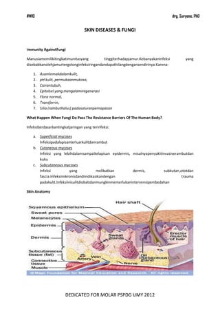
Skin Anatomy
DEDICATED FOR MOLAR PSPDG UMY 2012
2. #M10
drg. Suryono, PhD
Fungal Infections (Mycoses)
Principal tissue sites of deep mycoses in comparison to those of the superficial, cutaneous, and
subcutaneous mycoses
Portals
of
entry
of
pathogenic
and
opportunistic
fungi
causing
deep
mycoses
Superficial Mycoses
Disease
Etiological Agent
Symptoms
Identification Of Organism
Pityriasisve
rsicolor
Malassezia furfur
hypopigmente
d macules
"Spaghetti and meatballs" appearance of
organims in skin scrapings.
Tineanigra
Exophialawernecki
i
black macules
Black, 2-celled oval yeast in skin scrapings
Black
piedra
Piedraiahortai
black nodule
on hair shaft
black nodule on hair shaft composed of spore
sacs and spores
White
piedra
Trichosporombeig
elii
creme-colored
nodules
on
hair shaft
white nodule on hair shaft composed of
mycelia that fragment into arthrospores
DEDICATED FOR MOLAR PSPDG UMY 2012
3. #M10
drg. Suryono, PhD
PityriasisVersicolor
Keluhankulit yang umumdenganpenampilan patch yang tidakberwarnaterutamapada dada
danpunggung.
Istilah“pityriasis”digunakanuntukmendeskripsikankondisidimanaskala
patch
samaseperti bran. Sedangkan“versicolor”digunakanuntukmenggambarkanwarna-warna yang
muncul.Terkadangpenyakitinidisebut“Tineaversicolor”meskipun
kata
“tinea”secaralangsungdimaksudkanuntukinfeksidenganjamurdermatrofit.
Clinical Features
•
Pityriasisversicolormenyerangbagiantenggorokan,
leher,
dan/ataulengan,
jarangterdapatpadabagian
lain
tubuh.
Patch
berwarna
pink,
coklattembagaataulebihpucatdaripadakulit
di
sekitarnyadansedikitgatal.
Patch
pucatbiasanyapadakulit
yang
lebihgelap,
kondisiinidikenaldenganPityriasisversicoloralbadancenderungtidakgatal. Terkadang patch
bersisikdanberwarnacoklta, kemudianmenjaditidakbersisikdanberwarnaputih.
•
A yellow-green fluorescence dapatdiamatidenganWood's light(long wave ultraviolet A)
padadaerah yang terinfeksi.
•
Pityriasisversicolorseringterjadi di daerahpanas, beriklimlembabataupadaseseorang yang
berkeringatbanyak,
sehingga
bias
kambuhpadasetiapmusimpanas.
Pityriasisversicolortidakmunculpada area yang terkenasinarmataharilangsung.
Patch Putih
Persistent Pale Marks
Hypopigmented Macules
Black Nodule On Hair Shaft
Patch Coklat
DEDICATED FOR MOLAR PSPDG UMY 2012
4. #M10
drg. Suryono, PhD
Creme-Colored Nodules On
Hair Shaft
M. furfur
Superficial dermatophytes
M. furfur skin scraping with calcifluor stain.
Observe the detail of the fungal features with
this technique that stains the fungal elements
green-white.
Hypopigmentedtineaversicolor
Hypopigmentedtineaversicolor
Hypopigmentedtineaversicolor.
Small, pale macules coalesce into larger
patches on this patient's neck.
Glossary
•
Dermatophyte ; A type of fungus that causes diseases of the skin, including tinea or
ringworm.
•
KOH ;The chemical formula for potassium hydroxide, which is used to perform the KOH test.
The tests is also called a potassium hydroxide preparation.
•
Thrush ; A disease of the mouth, caused by and characterized by a whitish growth and
ulcers. It can be diagnosed with the KOH test.
DEDICATED FOR MOLAR PSPDG UMY 2012
5. #M10
•
drg. Suryono, PhD
Tinea ; A superficial infection of the skin, hair, or nails, caused by a fungus and commonly
known as ringworm.
Ring Worm
Ring Worm
Dermatomycosis (ringworm) of hair follicles
Patient with ringworm on the arm, or
Tineacorporis
due
to
Trichophytonmentagrophytes
Ringworm, stained preparation, Macroconidia
ofMicrosporumcanis
Ringworm,
stained
preparation,
Macroconidiaof Microsporumcanis
Cutaneous Mycoses
Infeksi yang lebihdalamkelapisan epidermis, contohnyapenyakit invasive rambutdan
kuku.Penyakitiniterbataspadalapisan keratin di kulit, rambut, dan kuku.Tidaksepertisuperficial
mycoses, host dariresponimundapatdiaktifkan, menghasilkanperubahanpatalogispadalapisan yang
lebihdalampadakulit.Organisme
yang
menyebabkanpenyakitiniadalahDermatophytes.PenyakitiniseringdisebutdenganingwormatauTinea.S
emuapenyakittersebutdisebabkanolehMicrosporum, Trichophyton, danEpidermophyton, yang
terdiridari 41 spesies.
DEDICATED FOR MOLAR PSPDG UMY 2012
6. #M10
drg. Suryono, PhD
Microsporum
Disease
Trichophyton
Symptoms
Epidermophyton
Identification Of Organism
Tineacapitis
Ringworm of scalp
Presence/absence and shape of
micro- and macroconidia in
scrapings of lesion, KOH mount
Ringworm of trunk, arms, legs
Presence/absence and shape of
micro- and macroconidia in
scrapings of lesion, KOH mount
Ringworm of hand
Presence/absence and shape of
micro- and macroconidia in
scrapings of lesion, KOH mount
Ringworm of groin "jock itch"
Presence/absence and shape of
micro- and macroconidia in
scrapings of lesion, KOH mount
Tineacorporis
Tineamanuum
Tineacruris
DEDICATED FOR MOLAR PSPDG UMY 2012
7. #M10
drg. Suryono, PhD
Tineapedis
Ringworm of foot "athlete's
foot
Presence/absence and shape of
micro- and macroconidia in
scrapings of lesion, KOH mount
Infection of nails
Presence/absence and shape of
micro- and macroconidia in
scrapings of lesion, KOH mount
Infection of hair shaft surface
Mycelium and spores on hair shaft
Infection of hair shaft interior
Mycelium and spores in hair shaft
Tineaunguium
Ectothrix
Endothrix
Subcutaneous Mycoses
Infeksiinimelibatkan
dermis,
jaringansubkutan,
otot,
fascia.Infeksiinikronisdandiindikasikandengan
padakulit.Infeksiinisulitdiobatidanmungkinmembutuhkanintervensipembedahan.
dan
trauma
Jenis-jenissubcutaneous mycoses:
1. Chromoblastomycosis
Ditandaidenganlesiverrucoidpadakulit
(biasanyapadaekstremitasbawah)
darihasilpemeriksaanhistologismenunjukkansel multiform (denganseptationstegaklurus)
disebutdengan"copper pennies" yang merupakancirikhasdariinfeksiini.
2. Mycetoma
Infeksisubkutansupuratifdangranulomatosamikosis yang merusaktulang contiguous,
tendon,
danototrangka.Mycetomadicirikandenganadanyasaluran
sinus
keringdaributirankecil yang berpigmentetapiterlihatjelassebagaibutiran yang ekstrusi.
3. Sporotrichosis
DEDICATED FOR MOLAR PSPDG UMY 2012
8. #M10
drg. Suryono, PhD
Disease
Etiological Agent
Symptoms
Id Of Organism
Nodules
and
ulcers
along
lymphatics and at
site of inoculation
Yeast in tissue; mold
at rm temp with
"rosette pattern"
Fonsecaeapedrosoi or
compacta,
Wangielladermatitidis
Warty
nodules
that progress to
"cauliflower-like"
appearance
a
inoculation site.
Copper-colored
spherical yeasts called
"Medlar bodies" in
tissue
Pseudallescheriaboydii,
Madurellagrisea
or
mycetomatis
Draining
sinus
tracts at site of
inoculation
White, brown, yellow
or black granules in
exudate that are
fungal colonies
Sporotrichosis
Sporothrixschenckii
1. yeast
2. mold
Chromoblastomycosis
Mycetoma
Systemic Mycoses
Infeksi
yang
berasaldariparu-parudandapatmenyebarkebanyak
system
organ.Organismeiniadalahorganismeinherenvirulen, kecualiCryptococcus adalahjamurdimorfik.
Opportunistic Mycoses
•
Infeksipadapasiendengandefisiensiimun, contohnya:
DM,terapi immunosuppressive, keganasan
•
Candidiasis (Candida albicancs)tumbuhsepertikrimpadapermukaantubuhcontohnya, mulut,
kulit, vagina. Budding yeast. Berbentukpseudohyphaepadajaringan, grem tube dalam serum
•
Aspergillosis(Aspergillusniger)
AIDS,
perubahan
DEDICATED FOR MOLAR PSPDG UMY 2012
flora
normal,
9. #M10
drg. Suryono, PhD
Oral Candidiasis
•
Synonyms: Candidosis, Thrush, Moniliasis
•
Infeksi opportunistic yang disebabkanolehjamurObiquitous, saprophytic dari genus Candida
yang mencakup 8 jenisjamur, yang paling umumCandida albicans
•
Candidiasis is usually limited to the skin and mucous membranes.
•
Common clinical types of mucocutaneous candidiasis include:
a.
b.
c.
d.
e.
•
oropharyngeal (affecting the oral cavity and/or pharynx)
vulvovaginal (affecting the vaginal and vulvar mucosa)
paronychial (affecting the nail beds and folds)
interdigital (usually affecting the skin in between the fingers)
intertriginous (affecting the skin of the submammary areas or the groin and/or scrotum)
infeksiCandidiasis,
sistemikdan
invasive
dapatterjadikhususnyapadapasiendengan
immunosuppression berat. Saluranpencernaan, trakea, paru-paru, hati, ginjal, dan system
sarafpusatberpotensialuntukterkenainfeksiCandidiasis
yang
nantinyaakannerakibat
septicemia, meningitis, hepatosplenic, dan endocarditis
Epidemiology
Oral candidiasis disebabkan palingdominanolehCandida albicans, meskipunCandidajenis lain
jugaterlibatpadainfeksitersebut.Candidamerupakanbagiandari flora normal mulutyaitusekitar30% 50%daripopulasi. Dan dapatmenyebabkaninfeksi opportunistic pada oral cavity denganberbagaijenis
factor predisposisi yang lain.
Etiology and Pathogenesis
Factor yang menyebabkanCandidiasis:
1. The immune status of the host
2. The oral mucosal environment
3. The particular strain ofCandida albicans (thehyphal form is usually associated with
pathogenic infection).
Specific Conditions That May Predispose A Patient To Develop Oral Candidiasis
1. Factors that alter the immune status of the host:
•
Blood dyscrasias or advanced malignancy
•
Old age/Infancy
•
Radiation therapy/Chemotherapy
•
HIV infection or other immunodeficiency disorders
•
Endocrine abnormalities:
DEDICATED FOR MOLAR PSPDG UMY 2012
10. #M10
drg. Suryono, PhD
•
Diabetes mellitus
•
Hypothyroidism or Hypoparathyroidism
•
Pregnancy
•
Corticosteroid therapy/Hypoadrenalism
2. Factors that alter the oral mucosal environment:
•
Xerostomia
•
Antibiotic therapy
•
Poor oral or denture hygiene
•
Malnutrition/Gastrointestinal malabsorption
•
Iron, folic acid, or vitamin deficiencies
•
Acidic saliva/Carbohydrate-rich diets
•
Heavy smoking
•
Oral epithelial dysplasia
Clinical Presentation AndTreatment
I. (Acute) Pseudomembranous Candidiasis
•
Pseudomembranous candidiasis is the most common form of oral candidiasis.
•
The most common sites include buccal mucosa, dorsal tongue, and palate.
•
Most frequent etiologies include antibiotic therapy or immunosuppression.
•
It appears as soft, creamy white to yellow, elevated plaques, that are easily wiped off
affected oral tissues and leave an erythematous, eroded, or ulcerated surface which may be
tender.
Rationale for Treatment: Topical vs. Systemic Drugs
•
Topical antifungals are usually the drug of choice for uncomplicated, localized candidiasis in
patients with normal immune function.
•
Systemic antifungals are usually indicated in cases of disseminated disease and/or in
immunocompromised patients.
•
Duration of therapy: Medication should be continued for at least 48 hours after the
disappearance of clinical signs of candidiasis along with complete healing and the absence of
mucosal erythema. Some sources recommend drug therapy should be continued for 10-14
days regardless of the disappearance of clinical signs of candidiasis.
DEDICATED FOR MOLAR PSPDG UMY 2012
11. #M10
drg. Suryono, PhD
II. Chronic Hyperplastic Candidiasis
•
The most common sites are the anterior buccal mucosa along the occlusal line, and
laterodorsal surfaces of the tongue.
•
The etiology may be idiopathic or associated with immunosuppression.
•
The most common appearance is that of asymptomatic white plaques or papules
(sometimes against an erythematous background) that are adherent and do not scrape off.
•
Some sources believe that hyperplastic candidiasis may have the ability to promote the
development of oral epithelial carcinogenesis.
Treatment of hyperplastic candidiasis:
•
Use topical or systemic medications as was recommended for pseudomembranous
candidiasis
III. Chronic Atrophic (Erythematous) Candidiasis
•
The most common site is the hard palate under a denture, but atrophic candidiasis may also
be found on the dorsal tongue and other mucosal surfaces.
•
The most common etiology is poor denture hygiene, and/or continuous denture insertion,
but it may also be caused by immunosuppression, xerostomia, or antibiotic therapy.
•
The most common appearance is that of a red patch or velvet textured plaque. When
atrophic candidiasis occurs on the hard palate in association with a denture, it is frequently
associated with papillary hyperplasia.
•
Patients may complain of a burning sensation associated with this type of candidiasis
•
It is important to remember to treat both the denture (if present) and the oral tissues. (The
denture will act as a reservoir for the Candida and reinfect the tissues if they are not treated
concurrently).
IV. Median Rhomboid Glossitis
•
Median rhomboid glossitis is a form of chronic atrophic candidiasis characterized by an
asymptomatic, elongated, erythematous patch of atrophic mucosa of the posterior middorsal surface of the tongue due to a chronic Candida infection. (In the past, median
rhomboid glossitis was thought to be a developmental defect resulting from a failure of the
tuberculumimpar to retract before fusion of the lateral processes of the tongue).
•
A concurrent "kissing lesion" of the palate is sometimes noted.
•
Specific predisposing etiologic factor(s) for median rhomboid glossitis have not been clearly
established.
DEDICATED FOR MOLAR PSPDG UMY 2012
12. #M10
drg. Suryono, PhD
V. Angular Cheilitis (Perleche)
•
Clinical appearance is that of red, eroded, fissured lesions which occur bilaterally in the
commissures of the lips and are frequently irritated and painful.
•
The most common etiology is loss of vertical occlusal dimension, but it may also be
associated with immunosuppression.
Diagnosis
•
The diagnosis of oral candidiasis is most frequently made on the basis of clinical appearance
along with exfoliative cytology examination. This involves the histologic examination of
intraoral scrapings which have been smeared microscope glass slides. A 10% - 20%
potassium hydroxide preparation ("KOH prep") can be used for immediate microscopic
identification of yeast cell forms. Alternatively, the slide containing the cytologic smear can
be sprayed with a cytologic fixative and stained using PAS (Periodic acid - Schiff) stain prior
to microscopic examination.
Candida
Alhamdulillah selesaijugangeditmateri
SUPER yang satuini.Maafyatemantemannggabisamaksimal translate
semuanya. Banyak (banget) kakaa –oa
Anyway, hope you guys can
understand the main point of this
Candidiasis
lecture
Oke,
adasatuquotenihdariomAristotelesyang
mungkinbisabikin kalian
lebihsemangat.
Here is …
“Pendidikanmempunyaiakar yang
pahit, tapibuahnyamanis”
SemangatHaphap!!
DEDICATED FOR MOLAR PSPDG UMY 2012