2. Jg college of nursing
Ahmedabad
Sub-Medical Surgical Nursing
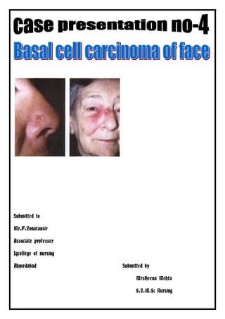
Topic- Basal cell carcinoma of face
Submitted to-Mr.P.Yonatansir
Submitted by-MrsHeena Mehta
Sr no content Page no
1 Identification data
2 History
3 Physical examination
4 Investigation
5 Disease condition
6 Definition
7 Pathophysiology
8 Management
9 Nursing diagnosis
10 Health teaching
11 Bibliography
3. IDENTIFICATION DATA
PATIENT’S NAME: JivatibenBhojabhaiGajjar
Indoor.no: F 56854
AGE:64 years
SEX:Female
DATE OF ADMISSION`:12-1-2012
DR’S UNIT: Unit-2 Dr.prakashpatel
WARD: cancer female medical ward
MARRITAL STATUS: married
RELIGION: Hindu
EDUCATION: Illiterate .
OCCUPATION: House wife
ADDRESS:Jetpur,Rajkot
DIAGNOSIS:Basal cell carcinoma of the face
HEIGHT: 146Cm
WEIGHT: 5 Kg
4. PRESENTING COMPLAINS:
Patient having complained of following:
-Fever
-Itching on face
-Black ulcer on the face
-Dryness of face skin
-Indigesion
-Weakness
PRESENT HISTORY:
Jivatiben has complain of the dryness of the face skin, itching of the face , black ulcer on the
face, loss of apitite and weakness since last 8 month so she has to gone for treatment at
private hospital in Jetpur (Rajkot) but symptoms not relieve itching constant occure than she
refer to the civil hospital for medication.
PAST HISTORY:
PAST MEDICAL HISTORY:
Upto 64 year she has lot off time taken medication for minor disease but not need any
hospitalization ,symptomatic medication taken for three to five day and symptoms relieve
but last 8 month she was suffering from the skin disease and symptoms not relieve thus refer
5. civil hospital and finally after total investigation she diagnose BASAL CELL CARCINOMA
OF THE FACE and admit in the civil hospital for treatment.
PAST SURGICAL HISTORY:
No any surgical treatment needed Jivatiben, No any surgery done to the Jivatiben.
DIET HISTORY:
Jivatiben taken normal diet in her life. She has a farm so her husband grow
normaly all types of vegetables in his farm .
PERSONAL HISTORY:
Diet : vegetarian & taking all type of small amount diet
Appetite : Decreased
Sleep :disturb
Micturation : No burning micturation
Bowel habit: Abnormal habits
Smoking : No
Alcohol : No
Drugs : No
Tobacco : No
No any other habits
FAMILY HISTORY:
In her family no any family members have history of any Hypertension, Diabetes mellitus,
Ischemic heart disease, Epilepsy, Asthma, Storks, Arthritis, Cancer or any other disease. Her
father suffering from the tuberculosis and expired with this disease.
Sr. Name of Family Age in Relationship
Education Occupation
No. Members Year With patient
1 BhojabhaiBudhabh 7oYrs. Husband Illiterated Farmer
6. aiGajjar
2 JivatibenGajjar 64Yrs Patient Illiterated Housewife
3 JentibhaiGajjar 46Yrs Son 8th pass Farmer
4 NandubenGajjar 40Yrs Son’s wife 7th pass Housewife
5 Ramesh Gajjar 20Yrs Grand son 12th pass Farmer
6 Neeta Gajjar Yrs Grand 10th -
daughter
7 RasikGajjar 25 son B.Com -
8 NanjiGajjar 20 son B.Com -
SOCIOECONOMIC HISTORY:
In Jivatiben’s house , her husband and elderson as a farmer so they work in farm and grow
seasional vegetables and cereals for family and for sale so her income is not fix sometime her
family earn more money and some times her family earn less money.
PHYSICAL EXAMINATION:
VITAL SIGN
Date Temp ( Pulse Respiration(/min) BP (mm of
F) (/min) Hg)
16-07- 100 F 100/min 20/min 118/74
2012
17-07- 99 F 126/min 24 min 122/64
2012
18-07- 99 F 120/min 26 min 114/78
2012
19-07- 98.6F 116/min 20 min 120/74
2012
GENERAL OBSERVATION:
Sensorium: She is conscious and well oriented
Foul body odour: no any bad odour from her body
Foul breath : no
Posture : normal
Hair: Brown hair, clean no any dandruff.
7. GENERAL APPERANCE:
Body image: normal
Health: Unhealthy
Activity: less active
MENTAL STATUS:
Consciousness: conscious
Look: weakness, fatigue due to her disease.
Posture
Body curves: normal
Movement: Full movement(if given deep pain than small reflection was done by
patient)
Height: 146cm Weight: 67kg
SKIN CONDITION:
Color: pallor
Texture: Rough skin
Temperature: warm
Lesions: no lesions present
HEAD & FACE:
Scalp: clean
Face: pale, fatigue, fear, anxiety
EYES:
Eyebrow: normal
Eye lashes: no infection, not open by patient
Eyelids: no any injury or oedema is present
Eye balls: not sunken
Conjunctiva: pale
Sclera: no jaundiced
Pupils: constricted
Vision: react to light
EAR:
External ear: no discharge present
8. Hearing: normal
NOSE:
External nares: Redness present
Nostrils: normal. keeping face mask for proper oxygenation
MOUTH & PHARYNX:
Lips: dry
odour of the mouth: not present
Teeth: normal
Mucus membrane: dry
Tongue: pale and moist
NECK:
Lymph node: Not palpable
Thyroid gland: normal
Range of motion: flexion, extension and rotation when done by someone, patient able
to done by own self.
CHEST:
Thorax: expansion
Breath sound: No any sound heard
Heart: normal
ABDOMEN:
Observation: no skin rashes and scar
Auscultation: reduced bowel sound
Palpation: no tenderness present
Percussion: not presence of gas, fluid or masses
EXTREMITIES:
Lower extremities: fully movements of lower extremities. mildoedema present
Upper extremities: can move both hands but mild oedema is present
Genital and rectum:
No enlarged inguinal lymph nodes, No hemorrhoids, no enlargement of prostate
glands.
Bladder & Bowel Pattern: Abnormal.
9. INVESTIGATION:
Serum Biochemistry test:
Investigation In patient Normal value
Hemoglobin 12 % gm% 14 – 17 gm %.
RBC 100 mg/dl 153mg/ml
UREA 24mg/dl 15-45mg/dl
WBC 9,200/cumm 4000-11000/cumm
S.creat. 0.59mg/dl 0.7-1.5mg/dl
SGPT 36U/L 0-55U/L
S. phosphate 108 U/L <50-150U/L
S.Billirubin 0.7mg/dl 0.2-1.2mg/dl
BLOOD CHEMISTERY
FASTING 90.0mg/ dl 70-110mg/dl
CHOLESTROL 174 mg/dl >240.0mg/dl
Serum Electrolytes:
Investigation In patient Normal value
S. Na+ 144.3 meq/L 135-145meq/L
S.K+ 4.62 meq/L 3.5-5.5 meq/L
Chloride 105 97-108
X-RAY CHEST:
Olcg in bothlungs
ECG:wnl
MEDICATION
CHEMOTHERAPY GIVEN AFTER SURGERY
-Injection 5FU and cyclophosphamide regimen every 3week for three cycle.
-Injection amikasine 500gm i/v 12hourly.
-Injection voveran 1 ampoule i/v 12hourly.
- Injection Ondensten 1 ampoule i/v sos
- Tablet-Rantac 150 mg 1 bd.
-Tablet- MV/BC 1 bd
10. Maintain intake and output chart daily
Contineus observation of the patient on monitor for any abnormal symptoms.
TPR chart 1 hourly Monitoring continuously for blood pressure, respiration rate,
pulse, and for oxygen saturation.
Care taken of catheter daily
Care taken of all tubes which are inserted
Watched for respiratory failure .
Changed the dressing and adhesive tap at the site of intracath.
DISEASE CONDITION
ANATOMY AND PHYSIOLOGY OF SKIN-
The skinis the largest organ in the body, comprising about 15% of the body weight. The
total skin surface of an adult ranges from 12 to 20 square feet. In terms of chemical
composition, the skin is about 70% water, 25% protein and 2% lipids. The remainder
includes trace minerals, nucleic acids, glycosoaminoglycans, proteoglycans and numerous
other chemicals.
11. The skin consists of three main layers: epidermis, dermis and subcaneous tissue.
The Epidermis
The epidermis is the topmost layer of the skin. It is the first barrier between you and the
outside world. The epidermis consists of three types of cells keratinocytes, melanocytes and
Langerhans cells. Keratinocytes, the cells that make the protien keratin, are the predominant
type of cells in the epidermis. The total thinkness of the epidermis is usually about 0.5 - 1
mm. At the lowermost portion of the epidermis are immature, rapidly dividing
keratinocytes. As they mature, keratinocytes lose water, flatten out and move upward.
Eventually, at the end of their life cylce, they reach the uppermost layer of the epidermis
called stratum corneum. Stratum corneum consists mainly of dead keratinocytes, hardened
proteins (keratins) and lipids, forming a protective crust. Dead cells from stratum corneum
continuously slough off and are replaced by new ones coming from below. The skin
completely renews itself every 3 - 5 weeks. Most mild peels work by partly removing the
stratum corneum and thus speeding up skin renewal.
Another significant group of cell in the epidermis are melanocytes, the cells producing
melanin, the pigment responsible for skin tone and color. Finally, Langerhans cells are
essentially a forepost of the immune system in the epidermis. They prevent unwanted
foreingn substances from penetrating the skin.
The condition of epdermis determines how "fresh" your skin looks and also how well your
skin absorbs and holds moisture. Wrinkles, however, are formed in lower layers.
The Dermis
The dermis is the middle layer of the skin located between the epidermis and subcutaneous
tissue. It is the thickest of the skin layers and comprises a tight, sturdy mesh of collagen and
elastrin fibers. Both collagen and elastin are critically important skin proteins: collagen is
responsible for the structural support and elastin for the resilience of the skin. The key type
of cells in the dermis is fibroblasts, which synthesize collagen, elastin and other structural
molecules. The proper function of fibroblasts is highly important for overall skin health.
The dermis also contains capillaries (tiny blood vessels) and lymph nodes (depots of
immune cells). The former are important for oxygenating and nourishing the skin, and the
latter -- for protecting it from invading microorganisms.
Finally, the dermis contains sebacious glands, sweat glands, hair follicles as well as a
relatively small number of nerve and muscle sells. Sebacious glands, located around hair
follicles, are of particular importance for skin health as they produce sebum, an oily
protective substance that lubricates and waterproofs the skin and hair. When sebacious
gland produce too little sebum, as is common in older people, the skin becomes excessively
dry and more prone to wrinkling. Conversely, overproduction or improper composition of
sebum, as is common in adolescents, often leads to acne.
The dermis is the layer responsible for the skin's structural integrity, elasticity and
resilience. Wrinkles arise and develop in the dermis. Therefore, an anti-wrinkle treatement
has a chance to succeed only if it can reach as deep as the dermis. Typical collagen and
elastin creams, for example, never reach the dermis because collagen and elastin molecules
are too large to penetrate the epidermis. Hence, contrary to what some manufacturers of
such creams might imply, these creams have little effect on skin wrinkles.
12. Subcutaneous tissue
Subcutanous (hypodermis) tissue is the innermost layer of the skin located under the dermis
and consisting mainly of fat. The predominant type of cells in the subcutaneous tissue is
adipocytes or fat cells. Subcutaneous fat acts as a shock absorber and heat insulator,
protecting underlying tissues from cold and mechanical trauma. Interestingly, most
mammals lack subcutaneous tissue because their fur serves as a shock absorber and heat
insulator. Sweat glands and minute muscles attached to hair follicles originate in
subcutaneous tissue.
The loss of subcutaneous tissue, often occurring with age, leads to facial sag and
accentuates wrinkles. A common procedure performed by dermatologists to counteract this
process is to inject fat (collected elsewhere in the body) under the wrinkles on the face .
DEFINITION:Basal cell carcinoma is the most common form of cancer worldwide and
accounts for about 80% of all cases of skin cancer.
CAUSES:
In Book In Patient
Radiation expose, UV Rays no
Gene Maturations May be
Arsenic exposure through ingestion no
Immunosuppression May be
Xerodermapigmentosum May be
Epidermodysplasticverruciformis no
Nevoid basal cell carcinoma syndrome No
Bazex syndrome No
Previous nonmelanoma skin cancer no
Rombo syndrome No
Alcohol consumption no
13. Types of basal cell carcinoma of the skin
In Book In My Patient
Nodular: About 60% of BCCs are nodular. There is small nodes seen over the face.
They start out as flat, well-defined lesions,
then often become small bumps, which
eventually collapse in the middle, leaving a
raised ring on the border. Most nodular
BCCs are on the face and so can be
disfiguring if not treated promptly.
Pigmented: Pigmented BCCs are similar to There is Black pigmentation seen over nodes
the nodular type, but they can have brown or
black spots in them. They can be confused
with some types of melanoma.
Fibrosing or Sclerotic: These BCCs are
usually found on the face and look similar to
scars. They are usually firm, ill-defined at the
border, flat or slightly depressed, yellowish
in color, and the surface tends to be smooth
and shiny.
Superficial: This type comprises about 15%
of BCCs. They spread outward from a red,
well-defined, scaly patch, most commonly
found on the trunk and limbs. They are easily
confused with psoriasis or eczema.
Fibroepithelioma of Pinkus: This is a rare
type of BCC. It tends to be a smooth,
elevated, small nodule found on the back,
extremities, groin, or sole of the foot. As
those are not sun-exposed areas, this disease
is probably not sun-related.
PATHOPHYSIOLOGY:
Over exposure to sun leads to the formation of thymine dimers, a form of DNA damage.
14. cumulative DNA damage leading to mutations.
Apart from the mutagenesis, over exposure to sunlight depresses the local immune system,
Basal-cell carcinoma also develops as a result of Basal-Cell Nevus Syndrome,
Developed tumors of the jaw, palmar or plantar (sole of the foot) pits, calcification of
the falxcerebri (in the center line of the brain) and rib abnormalities.
which inhibits the hedgehog signaling pathway.
A mutation in the SMO gene, which is also on the hedgehog pathway, also causes basal-cell
carcinoma
CLINICAL MENIFESTATION:
In Book In Patient
Present
skin bump or growth that is:
Pearly or waxy
White or light pink
Flesh-colored or brown
A skin sore that bleeds easily Present
A sore that does not heal Present
Oozing or crusting spots in a sore Not Present
Appearance of a scar-like sore without Present
having injured the area
Irregular blood vessels in or around the Not Present
spot
A sore with a depressed (sunken) area in Not Present
the middle
ASSESSMENT & DIAGNOSTIC FINDINGS:
15. IN BOOK IN PATIENT
- Taking a thorough history - Done
including family history
- Physical examination (note BP - Done
& weight)
- Laboratory work (cholesterol - Done
levels, glucose )
skin biopsy - Done
Shave biopsy uses a thin surgical
blade to shave off the top layers of
skin. This is the most common
method for diagnosing BCC.
Punch biopsy uses a round, cookie
cutter-like tool. It is used to take a
deeper skin sample.
MANAGEMENT:
Curettage and electrodessication
Surgical excision (removal)
Mohs surgery (also known as "micrographic surgery"), especially if the lesion is on the
face, is recurrent, has a diameter of greater than 2 cm, or is the sclerotic type
Topical creams such as imiquimod is FDA-approved for the treatment of superficial BCCs
not on the face, although studies have shown it can be effective against nodular BCC as
well
Excision
Simple surgical excision (removal) is used to treat both primary and recurrent tumors.
The procedure involves surgically removing the tumor and a certain amount of
normal-appearing skin surrounding it (the "margin"): For basal cell and squamous cell
carcinomas, margins are often 2 to 4 mm. The cure rates following excision are 95%
and 92% for primary BCC and SCC, respectively, and are dependent on the site, size,
and pattern of the tumor. Excision may be performed in the outpatient or inpatient
setting depending on the extent of the cancer.
Topical Creams
Since its approval in 2004, the immune systemactivator imiquimod (also known by the
brand nameAldara) has been a commonly prescribed topical (skin only) cream for
small superficial and nodular basal cell carcinomas, as well as a pre-cancerous
condition called actinic keratosis. It is spread on the lesion five times per week,
usually for six weeks, and completely clears the skin in about 88% of patients or more,
depending on the exact type of cancer. Another cream for superficial BCC is 5-
flourouracil (Carac or Efudex), a chemotherapy drug that is also used intravenously.
These treatments usually don't leave any scars, but they can cause considerable pain
and swelling as they work. Several other creams are being tested now,
16. including ingenolmebutate(PEP005), which is derived from a plant called the "petty
spurge."
Curettage and Electrodesiccation
Curettage and electrodesiccationis a simple, quick and effective method for destroying
small basal cell and squamous cell carcinomas. After scraping away the growth with a
long spoon-like instrument called a curette, the physician uses a mild electric current
to destroy any remaining abnormal cells. This scraping and cauterizing process is
typically repeated three times, and the wound tends to heal without stitches. It is best
for primary, not recurrent, lesions. The cure rates depend on the site: high-risk
locations (nose, ear, chin, mouth) have a recurrence rate of 4% to 18%, depending on
the tumor size. Recurrence rates decrease to 3% for tumors at low-risk sites of the
trunk and extremities. Overall, the 5-year cure rates for primary BCC and SCC treated
with C and E are 92% and 96%, respectively.
Mohs Surgery
TheMohs procedure (also known as Mohs micrographic surgery or margin controlled
excision) is an advanced technique developed in the 1940s by Dr. Frederic E. Mohsfor
removing lesions due to basal or squamous cell carcinoma. It involves removing thin
sections of the skin growth, layer by layer. Each layer is then examined under the
microscope, and removal of layers continues until no cancerous cells remain. It has the
highest cure rate of any skin cancer treatment and doesn't cause as much scarring as
other methods. It is especially useful for treating recurring skin cancer, larger tumors,
tumors on the ear, eyelid, nose, lip, or hand, tumors in sites prone to recurrence, and
the sclerotic subtype of basal cell carcinoma. It is the "gold standard" treatment: The
5-year recurrence rate is 1% for BCC and 3% for SCC. However, it is more costly,
time-consuming, and labor-intensive than other methods.
MEDICAL MANAGEMENT:
- Chemotherapy in four cycle with Adreamycine andcyclophosphemide.
- Analgesics
- Antibiotics
NURSING MANAGEMENT:
- Identify at risk patients, & teach lifestyle modifications to prevent development any
complication.
- Teach patient to control cholesterol levels through dietary reduction of cholesterol
intake, exercise, smoking cessation.
- Note & report findings from history, physical examination, & laboratory results that
indicate hypertension or diabetes, &teach to control blood pressure by taking
treatment in the nearest hospital.
NURSING DIAGNOSIS:
17. 1. Risk for infection related to decrease immune system.
2. Altered body temperature due to presence of infection.
3. Imbalance nutritional level less than body requirement related to loss of appetite.
4. Activity intolerance related to disease.
5 Impaired body image due disease.
6 Altered self image and confidence due to fegure.
HEALTH TEACHING:
Explain patient’s relatives about discharge planning.
Give advice about regular medication as per timing.
Advice given about good nutritive diet.
Advice given for prevention of infection management.
Prevention
The best way to prevent skin cancer is to reduce your exposure to sunlight. Ultraviolet light
is most intense at midday, so try to avoid sun exposure during these hours. Protect the skin
by wearing hats, long-sleeved shirts, long skirts, or pants.
Always use sunscreen:
Apply high-quality sunscreens with SPF (sun protection factor) ratings of at least 15.
Look for sunscreens that block both UVA and UVB light.
Apply sunscreen at least 30 minutes before going outside, and reapply it frequently.
Use sunscreen in winter, too.
Possible Complications
Untreated, basal cell cancer can spread to nearby tissues or structures, causing damage.
This is most worrisome around the nose, eyes, and ears.
Explain about follow up care.
18. BIBLIOGRAPHY:
1. Bennette and Plum; “TEXTBOOK OF MEDITION ; 10thedition, 1996;
W.B. Saunders Company, New York : 1996. PP :
2. Black J.M; “MEDICAL SURGICAL NURSING; 5th edition, 1999
; W.B. Saunders Company, Philadelphia. PP:
3. Brunners&Suddarth’s; “TEXT BOOK OF MEDICAL SURGICAL
NURSING VOL-_1”;10th edition, 2004; Elsevier Publishers, New Delhi,
India. PP:
4. B T Basavanthappa;”TEXT BOOK OF NURSING THEORIES”,Jaypee brothers
Medical Publishers ,New Delhi.
PP: 40-
WEBSITES:
- http://www.wikipedia.com.
- http://www.patho.skindisease.org/.com.in
- http://www.google.com.
- http://www.medicine.com.